What is retinal vein occlusion?
Retinal vein occlusion is due to the partial or complete blockage of the veins responsible for draining blood from the retina. The retina plays a crucial role in vision by converting light that enters the eye into nerve impulse signals, which are then sent to the brain to form the images we see. When these retinal veins become obstructed, it can lead to retinal swelling and increased pressure within the eye, potentially causing significant vision loss. Prompt treatment is essential to prevent or minimize vision damage.
Types of retinal vein occlusion
- Central retinal vein occlusion (CRVO)
- Branch retinal vein occlusion (BRVO)
How prevalent is retinal vein occlusion?
Retinal vein occlusion is the second most prevalent retinal vascular disorder after diabetic retinopathy.
- Retinal vein occlusion affects approximately 16 million people worldwide.
- Central retinal vein occlusion occurs in about 1-4 people per 1,000.
- Branch retinal vein occlusion occurs in about 6-12 people per 1,000.
What are the symptoms of retinal vein occlusion?
Symptoms of retinal vein occlusion typically occur in one eye.
- Blurred vision or sudden vision loss, which may develop rapidly or gradually over 2-3 hours or 2-3 days
- Floaters, such as lines or dark spots
- Eye pain, usually in severe cases

What causes retinal vein occlusion?
When the retinal artery becomes stiff from aging or plaque buildup, it can exert pressure on the retinal vein. This pressure may damage the retinal vein lining, leading to the formation of blood clots. Blood clots and slow blood flow can lead to retinal vein occlusion.
What are the risk factors of retinal vein occlusion?
- Age over 40 years. Retinal vein occlusion is most common in people aged 50-60, but it can affect people in their 40s.
- Diabetes
- Glaucoma
- Hypertension
- Atherosclerosis
What are the complications of retinal vein occlusion?
- Cystoid macular edema, resulting in blurry vision or vision loss
- Neovascularization of the eye, often occurring in the iris, is found in about 25% of patients
- Vitreous hemorrhage, which is caused by fragile new blood vessels that easily rupture
- Neovascular glaucoma, where abnormal blood vessels can increase eye pressure
- Retinal detachment.
Patients are also at higher risk of coronary artery disease and stroke due to hypertension and atherosclerosis.
How is retinal vein occlusion diagnosed?
Eye examination
An ophthalmologist will dilate the pupil and examine the back of the eye for:
- Vision loss
- Blockage in the central or branch retinal veins
- Macular edema
- New blood vessel growth
- Retinal ischemia
- Retinal hemorrhage
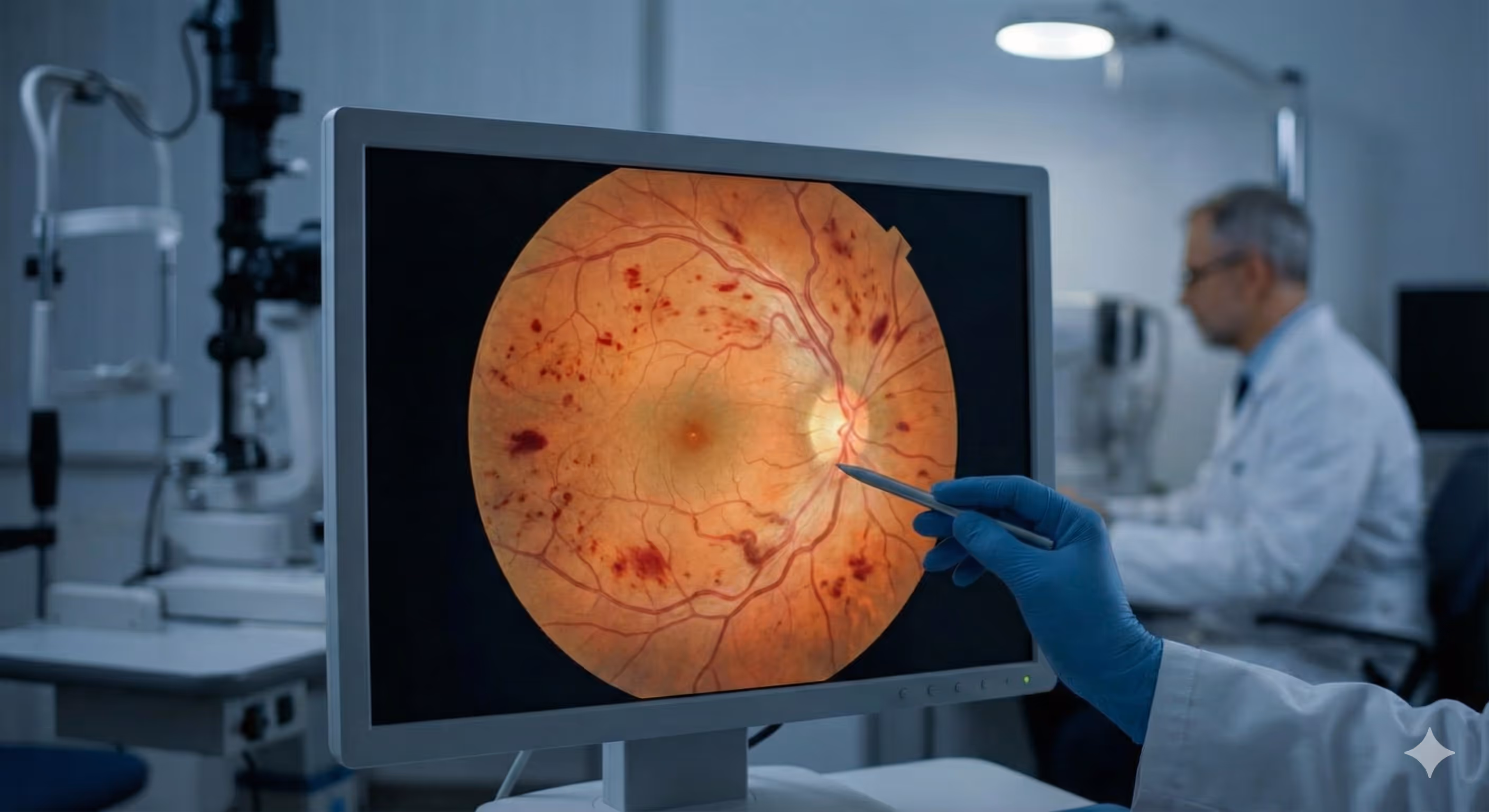
Fundus photography
Fundus photography to assess the presence of new blood vessels and evaluate the extent of bleeding in the eye.
Optical coherence tomography
Optical coherence tomography to check for macular edema and measure retinal thickness
Fluorescein angiography
Fluorescein angiography: A dye is injected into a vein to assess blood flow to the retina.
Additionally, the ophthalmologist may advise blood tests to evaluate cholesterol and blood glucose levels for further assessment.

How is retinal vein occlusion treated?
Currently, there is no cure for retinal vein occlusion. Treatment focuses on preventing the progression of symptoms, managing complications, and controlling risk factors.
- Anti-VEGF injections are the first-line treatment for patients with macular edema. VEGF (Vascular Endothelial Growth Factor) is a protein that stimulates the growth of blood vessels; increased secretion from the ischemic retina can cause the formation of new, fragile blood vessels that are prone to rupture. Before administering the injection, the doctor will apply an anesthetic eye drop to numb the eye. Patients may need several injections over the first 1–2 years, depending on the severity of their condition. The medications used for this treatment include Faricimab, Aflibercept, Bevacizumab, and Ranibizumab.
- Steroid injections to reduce swelling; however, these injections may increase the risk of cataracts and elevated intraocular pressure. Therefore, doctors typically recommend anti-VEGF injections as the first line of treatment.
- Panretinal Photocoagulation (PRP) is a laser eye treatment used to reduce the levels of VEGF protein, which is responsible for the formation of new, fragile blood vessels and bleeding in the eye. This procedure helps maintain normal intraocular pressure.
- Pars Plana Vitrectomy (PPV) is a surgical procedure recommended for patients with retinal detachment or those who have experienced persistent vitreous hemorrhage for more than four weeks or recurrent bleeding within the eye.
- Medications to control blood pressure, cholesterol, or diabetes, as these conditions increase the risk of new blood vessel formation.
How can I prevent retinal vein occlusion?
- Eat a balanced diet from all five food groups, emphasizing fruits and vegetables high in antioxidants to support vascular health.
- Engage in at least 30 minutes of exercise daily to enhance blood circulation and lower the risk of blood vessel blockages.
- Maintain a healthy weight.
- Quit smoking.
- Get annual health check-ups to assess risks and detect problems early.
What are self-care tips for patients with retinal vein occlusion?
Patients with retinal vein occlusion may feel stressed due to frequent doctor visits and treatments. They may also face driving restrictions during treatment. Patients experiencing stress or distress should consult their doctor or connect with others facing similar challenges to share experiences and learn coping strategies.
Questions you may consider asking your doctor
- What caused the retinal vein occlusion?
- Will the retinal vein occlusion affect my vision?
- What treatment methods are available, and what are their advantages and disadvantages?
A note from MedPark’s doctors
When discussing blood vessels, most people rarely consider those in the retina. Despite their small size, these vessels play a crucial role in vision. Blockages in these vessels can lead to severe vision problems. Prioritizing eye health and scheduling regular eye exams can help detect issues like retinal vein occlusion early, preventing potential complications.