





Choose the content to read
- What is an intravitreal injection?
- Why intravitreal injections?
- What conditions are amenable to intravitreal injections?
- How is an intravitreal injection performed?
- How safe is intravitreal injection?
- Intravitreal injections and long-term visual outcomes
- Frequently Asked Questions (FAQ) About Intravitreal Injections
- A note from MedPark’s doctors
Intravitreal Injection
An intravitreal injection is a treatment for various retinal and ocular vascular conditions, such as wet age-related macular degeneration and diabetic retinopathy. It helps slow the progression of vision loss and, in some cases, may improve visual acuity. It is a brief outpatient procedure, and hospitalization is typically not required. The frequency and number of injections are individualized to each patient, depending on the nature of the disease and the response to treatment.
What is vitreous humor?
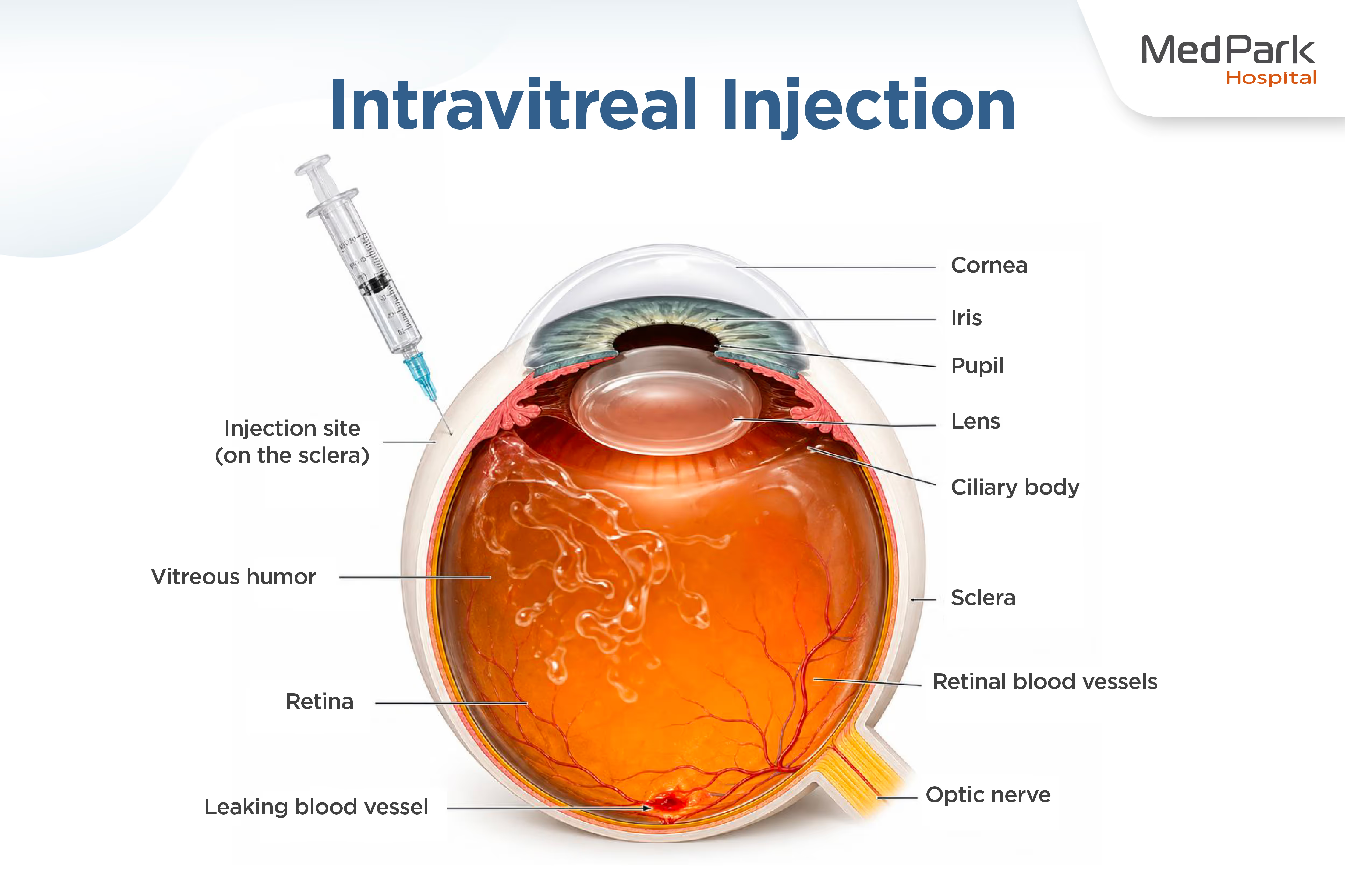
The vitreous humor is a clear, gel-like substance, similar to egg white, that fills the space inside the eye. It helps maintain the shape of the eyeball and accounts for approximately 80% of the eye’s total volume. With aging, the vitreous gradually degenerates from a firm gel to a more liquefied state. This change may lead to symptoms such as seeing floaters and flashes of light. In some cases, the vitreous may detach, potentially affecting the retina.
What is an intravitreal injection?
An intravitreal injection involves a doctor injecting medication through the sclera (the white part of the eye) into the vitreous humor for direct drug delivery to the retina. It is a treatment for retinal diseases that affect vision.
Why intravitreal injections?
- Targeted delivery to the site of disease
An intravitreal injection allows medication to reach the affected area directly. Many retinal conditions, such as age-related macular degeneration and diabetic retinopathy, occur at the level of the retina. Delivering medication into the vitreous humor enables it to act precisely at the site of disease for immediate therapeutic effect. - Blood–retinal barrier
The eye has a specialized barrier between the bloodstream and the retina that helps maintain a stable internal environment and prevents toxins or foreign substances from entering and damaging ocular tissues. As a result, medications taken orally or administered intravenously have limited ability to reach the site of disease within the eye. - Fewer systemic side effects
An intravitreal injection involves local delivery of a small amount of medication directly into the eye, minimizing systemic absorption and reducing the risk to cardiovascular or hematologic systems.
What conditions are amenable to intravitreal injections?
- Diabetic retinopathy: The injection treats diabetic macular edema and proliferative diabetic retinopathy caused by abnormal neovascularization by inhibiting the growth of new vessels and decreasing fluid leakage.
- Age-related macular degeneration: Particularly the wet form, characterized by abnormal blood vessel growth beneath the retina.
- Retinal vein occlusion: Treats macular edema that develops following blockage of a retinal vein; if left untreated, this may lead to permanent vision loss.
- Retinal conditions associated with leakage or swelling: For example, macular edema after cataract surgery, uveitis, or intraocular infections
However, an intravitreal injection is not suitable for every patient. The choice of treatment depends on severity and the ophthalmologist’s discretion. Key considerations include:
- Disease stage: In patients with early or moderate diabetic retinopathy, careful observation combined with optimal glycemic control may be preferred over immediate initiation of intravitreal therapy.
- Visual acuity: For patients with diabetic macular edema who retain good visual acuity (20/25 or better), doctors may opt for close observation before considering intravitreal injections
- Individualized treatment approach: Some patients may respond well to alternative modalities, such as laser therapy or surgical intervention, depending on lesion characteristics and patient-specific factors
How is an intravitreal injection performed?
Intravitreal injection is a minimally invasive and safe, brief outpatient procedure. It is performed under strict aseptic conditions in a carefully controlled environment to ensure patient safety and minimize the risk of infection.
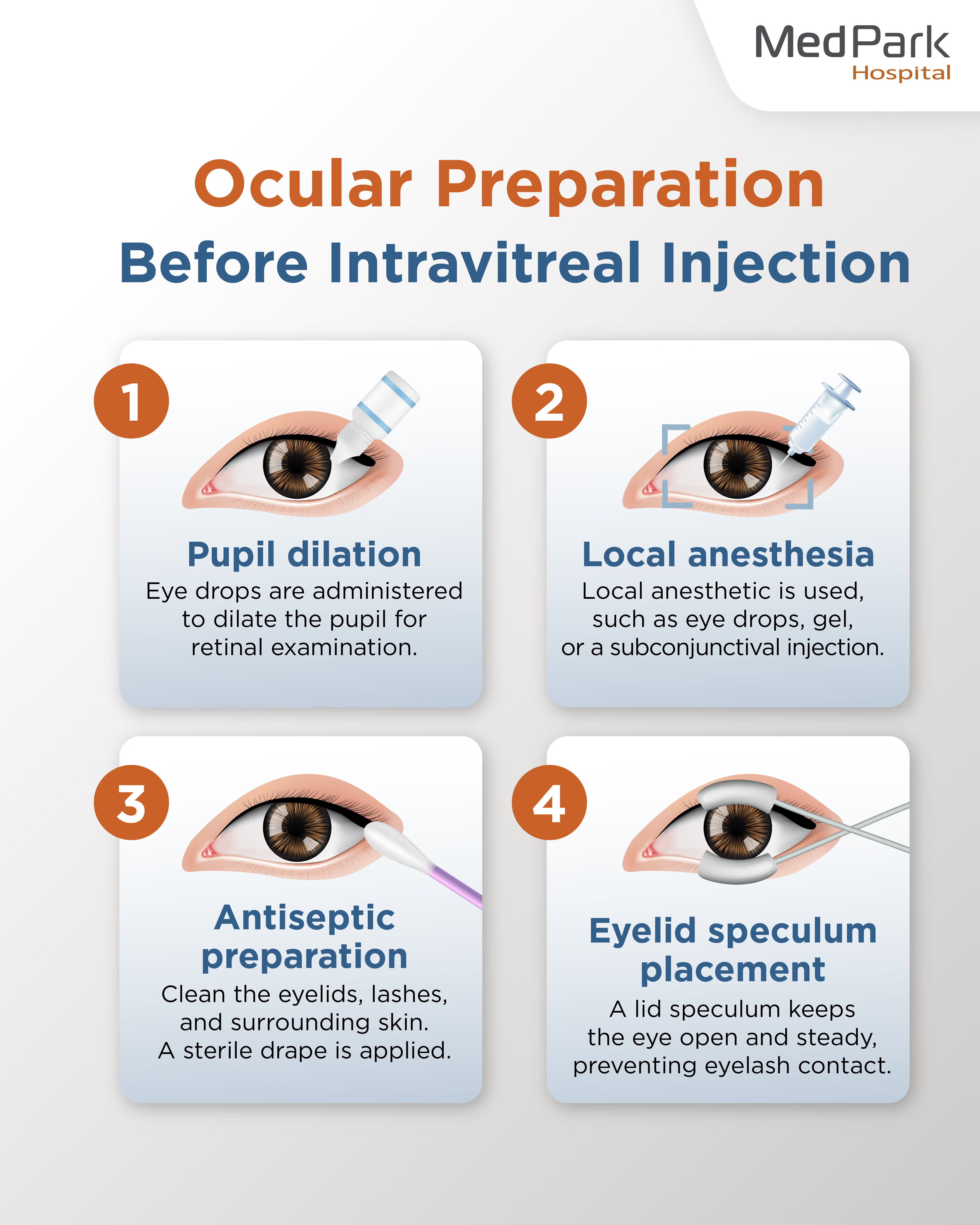
Ocular preparation
- Pupil dilation: The nurse or ophthalmologist administers pupil-dilating eye drops to facilitate a thorough retinal examination.
- Anesthesia: Anesthetic eye drops, gel, or a subconjunctival injection provide local anesthesia to ensure patient comfort throughout the procedure.
- Antiseptic preparation: Antiseptic solution disinfects the eyelids, eyelashes, and periocular skin. Staff places a sterile drape on the patient’s face.
- Eyelid speculum placement: Doctors place a lid speculum to keep the eye open, prevent eyelash contact with the injection site, and ensure ocular stability during the procedure
Intravitreal injection procedure
- The ophthalmologist carefully identifies the injection site on the sclera, typically located approximately 3.5–4 mm posterior to the limbus.
- Doctors instruct the patient to gaze in the opposite direction from the injection site and swiftly use a very fine needle to deliver the medication precisely into the mid-vitreous region.
- Following the procedure, patients may experience mild pressure or a transient sensation of foreign body in the eye.
Post-injection care
The ophthalmologist may gently apply sterile cotton to the injection site to minimize backing up of injected medication and remove any residual antiseptic solution.
- Most patients can resume normal daily activities on the same day.
- Administer antibiotic eye drops as directed to reduce the risk of inflammation and infection.
Post-injection precautions to prevent infection and contamination
- Do not rub the treated eye to prevent the risk of infection and wound dehiscence.
- Avoid wetting the treated eye for at least 3 days. When washing your face, use a clean, damp cloth instead. Refrain from swimming to reduce the risk of infection from pool water or natural bodies of water.
Temporary side effects may occur following the injection and typically resolve spontaneously within 1–2 weeks.
- A small red spot on the white of the eye caused by the injection needle is harmless and gradually resolves on its own.
- A gritty sensation, tightness, or pressure in the eye due to transient pressure during the injection; some patients may also experience mild irritation from the antiseptic used before the procedure.
- The appearance of black spots or shadows may result from small air bubbles introduced during the injection or residual medication particles in the vitreous. These typically resolve as the bubbles dissipate or the medication dissolves.
What post-injection signs and symptoms require urgent medical attention?
- Severe or progressively worsening eye pain
- Sudden or worsening vision loss
- Marked eye redness or purulent (pus-like) discharge
- Significant light sensitivity (photophobia)
- New onset of flashes of light or a rapid increase in floaters
How safe is intravitreal injection?
An intravitreal injection is a highly safe treatment, with serious complications occurring only rarely when performed by an experienced ophthalmologist under sterile conditions.
- Endophthalmitis: The risk of intraocular infection is extremely low, estimated at approximately 0.019% to 0.09%
- Retinal detachment: A rare complication, with an incidence of approximately 0.077% per injection
- Subconjunctival hemorrhage: Occurs in about 10% of patients, presenting as a localized red patch on the white of the eye. It is innocuous and typically resolves within 1–2 weeks.
- Transient elevation of intraocular pressure: A temporary rise in intraocular pressure may occur following the procedure.
How many intravitreal injections are required? Is a single injection sufficient?
Intravitreal injection therapy is not a one-time, curative treatment. Most retinal diseases managed with this approach are chronic conditions that require ongoing treatment, regular follow-up, and repeated injections to control disease activity and slow the progression of vision loss.
Why do I need multiple intravitreal injections?
- Short duration of therapeutic effect: Anti-VEGF agents have a relatively short duration of action. After injection, their therapeutic effect gradually wanes.
- Ongoing production of disease mediators: The treatment does not curtail the production of vascular endothelial growth factor (VEGF), a protein that promotes abnormal blood vessel growth.
- Risk of disease recurrence with treatment interruption: Patients who receive injections irregularly or discontinue treatment prematurely are at significantly increased risk of permanent vision loss due to disease recrudescence.
Ophthalmologists usually divide treatment into two main phases:
- Loading phase: To resolve fluid leakage and achieve a dry macula, intravitreal injections are administered monthly for a minimum of three doses.
- Maintenance phase: Treatment is then individualized based on the patient’s response.
- Pro re nata (PRN) regimen: Patients undergo regular monitoring, typically with monthly follow-up visits. Doctors administer the injection as needed when the disease recurs, such as recurrent macular edema or new hemorrhage.
- Treat-and-Extend (T&E) regimen: This is a widely adopted strategy. Doctors may stretch the follow-up interval from 4 to 6 or 8 weeks, and in some cases up to 12–16 weeks. This approach reduces treatment burden while maintaining effective disease control and minimizing the risk of recurrence.
Treatment monitoring and follow-up
Patients should adhere to scheduled follow-up visits to ascertain therapeutic response and visual outcomes.
- Imaging assessment: Doctors routinely order optical coherence tomography (OCT) to detect residual or recurrent retinal fluid, even when patients report improvement in vision.
- Individual variability: Some patients may be able to discontinue injections after a period of stability, but many require lifelong therapies to preserve vision.
- Innovations to reduce treatment burden: Longer-acting agents are available to extend a dosing interval. For example, Faricimab allows injection intervals of up to approximately four months. In addition, implantable, refillable intraocular drug delivery systems, such as the Port Delivery System, help reduce the frequency of hospital visits.

Intravitreal injections and long-term visual outcomes
The primary goal of intravitreal injection therapy is to suppress disease activity and slow progression, thereby preserving visual function for as long as possible. These agents work by reducing vascular leakage and inhibiting abnormal neovascularization, which are the key mechanisms underlying macular edema and retinal hemorrhage. With effective treatment, many patients can continue daily activities such as reading and driving.
Retinal diseases managed with intravitreal injections are chronic conditions requiring long-term care, rather than one-time, curative treatment. Failure to adhere to follow-up appointments or prolonged intervals between injections may lead to recurrent fluid accumulation, the development of fibrosis or irreversible retinal atrophy, and ultimately permanent vision loss that is not restorable.
Variability in treatment outcomes
The response to intravitreal injection therapy varies among individuals, influenced by several patient-specific factors:
- Baseline visual acuity: Patients with poorer baseline vision (e.g., 20/50 or worse) may demonstrate more substantial gains in visual acuity with appropriate treatment. For those with relatively good initial vision, maintaining disease stability and preventing further deterioration are primary management objectives.
- Ocular characteristics: Anatomical and physiological factors, such as axial length, age-related vitreous liquefaction, and fluctuations in intraocular pressure, can affect intraocular drug distribution and the duration of therapeutic effect.
- Pre-existing conditions and treatment response: Glycemic control and blood pressure can affect treatment outcomes. And some patients may respond more favorably to one agent than another.
Frequently Asked Questions (FAQ) About Intravitreal Injections
- Q: Is an intravitreal injection painful? Is it safe?
A: Intravitreal injections are generally not as painful as many patients expect. Before the procedure, the ophthalmologist administers topical anesthesia to numb the eye. Patients may experience a mild sensation of pressure or tightness during the injection. After the anesthetic wears off, some may notice mild irritation, a foreign body sensation, or dull discomfort, which typically resolves spontaneously within 24 hours. - Q: How much does an intravitreal injection cost?
A: The cost of treatment varies depending on the type of medication used, as well as other factors such as treatment regimen and healthcare facility. - Q: Can frequent intravitreal injections cause long-term harm to the eye?
A: Although intravitreal injections are generally considered safe, some studies suggest that receiving more than seven injections per year may be associated with an increased risk of glaucoma. Transient elevations in intraocular pressure may affect blood flow to the retina and optic nerve. Therefore, ophthalmologists carefully monitor intraocular pressure and retinal status before and after each treatment. - Q: Can intravitreal injections be discontinued once symptoms improve?
A: Some patients may be able to discontinue injections after a period of treatment. However, for those with chronic retinal conditions, such as age-related macular degeneration or retinal vein occlusion, ongoing therapy is often required to maintain visual function. - Q: What happens if intravitreal injections are delayed or missed?
A: Without timely and appropriate treatment, there is a significant risk of permanent vision loss. Persistent fluid accumulation within the retina can damage photoreceptor cells, leading to fibrosis or retinal atrophy, both of which are irreversible. - Q: Will vision improve immediately after an intravitreal injection?
A: Intravitreal injections do not result in immediate visual improvement. One to two days after the procedure, patients may experience mild blurred vision due to irritation from the antiseptic solution or notice floaters, typically caused by small air bubbles or residual medication particles within the vitreous. These symptoms usually resolve within 1–2 weeks. Vision typically improves after a series of injections.
A note from MedPark’s doctors
Intravitreal injection therapy is for managing chronic eye conditions. It is not a one-time cure but requires ongoing, consistent treatment in accordance with the physician’s recommended plan. Adherence to treatment helps control disease activity, slow retinal degeneration, and reduce the risk of permanent vision loss. Even in patients who have mild symptoms or relatively good vision, inadequate treatment or poor follow-up may allow the disease to progress silently.
Therefore, regular monitoring and strict adherence to the prescribed treatment regimen are essential to preserving long-term visual function.