





What is a Brain MRI?
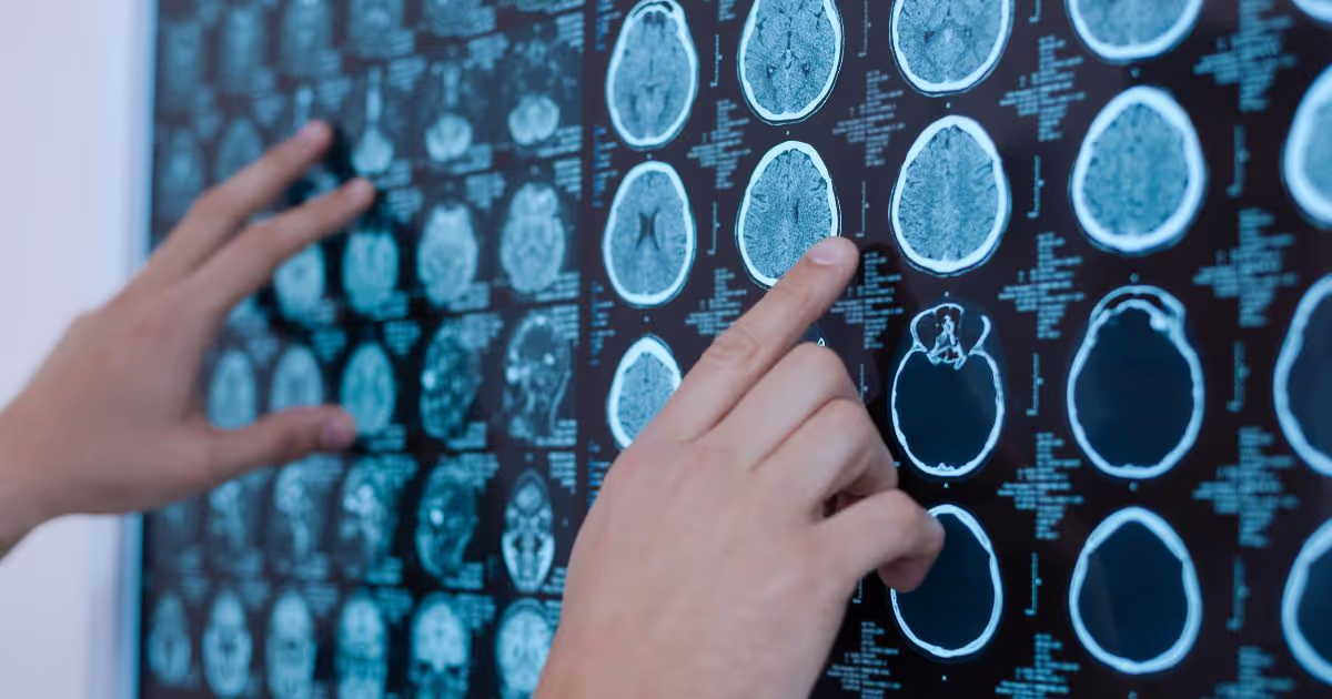
A brain MRI (Magnetic Resonance Imaging) is an advanced imaging technique that uses a powerful magnetic field and radio waves to create detailed, three-dimensional images of the brain and structures of the head. This technology enables doctors to evaluate internal anatomy in detail.
What can a brain MRI visualize?
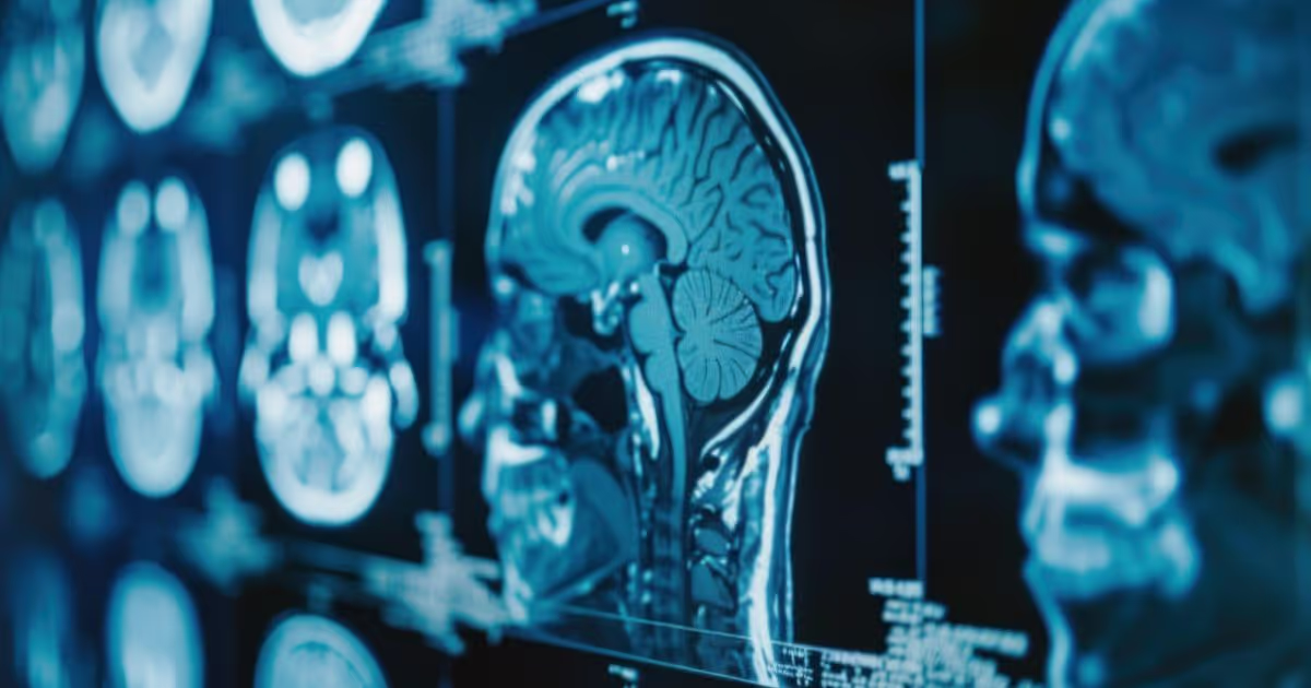
- The entire structure of the brain, including the cerebrum, cerebellum, and brainstem.
- Cerebral blood vessels and arteries.
- The eyes, extraocular muscles, optic nerves, and inner ear structures.
- Major cranial nerves that control facial movement, sensation, and balance.
- The skull and facial bones.
- Structures at the base of the skull, including muscles, fat, and connective tissues.
What conditions can a brain MRI detect?
- Tumors, cysts, or abnormal lesions
- Intracranial hemorrhage or stroke
- Brain swelling (edema), encephalitis, or meningitis
- White matter diseases
- Hydrocephalus (excess cerebrospinal fluid in the brain)
- Structural abnormalities of the brain and skull
- Head and brain injuries
What is a brain MRI with contrast?
A brain MRI with contrast is an imaging procedure that involves the intravenous administration of a gadolinium-based contrast agent before or during the scan. This contrast enhances image clarity and detail, allowing for more precise visualization of brain structures. Gadolinium temporarily alters the magnetic properties of water molecules in the body, causing certain tissues to appear brighter on the images and improving diagnostic accuracy.
Why is a contrast agent used?
The contrast agent travels through the bloodstream and tends to accumulate in areas of abnormality, making these lesions more visible on imaging.
- Helps delineate tumor boundaries and assess their blood supply.
- Aids in identifying inflammation and infections, such as brain abscesses or meningitis, which typically show increased contrast uptake.
- Evaluates lesions, such as in multiple sclerosis, to determine whether they are actively inflamed.
- Enhances visualization of blood vessels, helping detect narrowing or blockages in cerebral circulation.
What conditions can a brain MRI with contrast help detect?
- Dementia: Helps rule out other underlying causes of cognitive decline.
- Encephalitis: Clearly highlights areas of brain inflammation.
- Meningitis: Visualizes the extent of inflammation in the meninges and spine.
- Brain abscess: Helps define the abscess boundary more clearly.
- Multiple sclerosis: Differentiates active inflammatory lesions from old scars.
- Stroke: Assesses brain parenchymal tissue damage and evaluates blood flow.
- Tumor and brain metastasis
Who performs a brain MRI?
Radiology technologist
The radiologic technologist oversees the entire scanning process from start to finish. These include conducting thorough safety screening to ensure that no ferromagnetic objects enter the MRI room, positioning the patient on the examination table, operating the MRI machine, and verifying that the images obtained are clear and of high diagnostic quality.
Radiologist
A radiologist is a doctor specializing in interpreting medical imaging. They meticulously analyze the MRI images to detect abnormalities such as tumors, lesions, or vascular conditions, and compile their findings into a formal radiological report. This report is then shared with the primary doctor to provide an accurate diagnosis and appropriate treatment planning.
Are there any risks associated with a brain MRI?
A brain MRI is generally a safe procedure. However, one should be aware of certain risks:
1. Metallic devices and objects
The most significant risk is the powerful, always-on magnet of the MRI machine, which attracts ferromagnetic metal objects and interferes with the function of electronic devices in the body.
- Implanted devices, such as pacemakers and cochlear implants, may malfunction or stop working during the scan.
- Metal inside the body, including stents, surgical staples, prosthetic joints, artificial heart valves, aneurysm clips, shrapnel, or even tiny metal fragments in the eye, may shift or heat up, potentially causing tissue damage.
- External ferromagnetic metallic objects, such as earrings, necklaces, or clothing with metal frames, will fly like a projectile toward the machine or cause burns.
2. Adverse reactions to contrast agents
Although MRI contrast agents are generally very safe, some patients may experience side effects, including:
- Allergic reactions, such as hives, itching, or red/irritated eyes
- General discomfort, including nausea, abdominal discomfort, or dizziness
- Nephrogenic Systemic Fibrosis (NSF) may occur in patients with kidney disease when they cannot rapidly eliminate the contrast agent. It can lead to thickening and stiffening of the skin and internal organs.
3. Environmental factors during the scan
- The MRI machine has a narrow, tunnel-like gantry, which may elicit feelings of discomfort, anxiety, or panic in some individuals, especially those with claustrophobia.
- The machine produces intermittent rhythmic loud noises during operation, which may be disturbing or temporarily affect hearing.
- Radiofrequency energy may cause a slight increase in body temperature, which is usual. However, in some cases, such as in individuals with tattoos or medicated skin patches, localized heat buildup may occur, potentially leading to skin irritation or minor burns.
4. Pregnancy
Although there is no clear evidence that MRI is harmful to a fetus, it is generally advisable to forgo the scan during the first trimester unless necessary. One should avoid using contrast agents in pregnancy.

How should I prepare for a brain MRI?
Before the procedure:
- If you are claustrophobic or harbor any metal implants in your body, such as a pacemaker or metallic fragments from an injury, inform your doctor in advance.
- Continue taking your regular medications unless your doctor advises you to stop them temporarily.
- You may eat and drink as usual; however, you should check with your doctor whether you need to limit caffeine or alcohol intake.
- On the day of the exam, avoid wearing makeup, as some cosmetics may contain metallic particles. Also, do not wear jewelry or clothing with metal components, such as underwire bras.
- Change into a hospital gown provided by the facility.
During the procedure:
- You will lie supine on a motorized table that slides into the MRI scanner. A specialized head coil, similar to a helmet, will be positioned around your head, and you will receive earplugs or headphones to dampen the noise.
- You must be completely still throughout the scan (approximately 20–45 minutes), as movement can blur image quality and may necessitate a repeat scan.
- If contrast is required, a nurse will start an IV line in your arm or hand. You may feel a brief warm flushing in your face during the contrast injection; this is common and temporary.
After the procedure:
- You can return to your normal daily activities immediately after the scan.
- If you were given a sedative before the procedure, you should have someone accompany you or drive you home, as the effects may last for several hours.
- If you receive contrast, drink plenty of water to help your kidneys excrete it from your body more expeditiously.
- A radiologist will analyze the images and write up a report for your attending doctor, who will then contact you to explain the results and discuss the next steps in your care.

Why Choose Brain MRI at MedPark Hospital?
Brain health should never be overlooked, especially when experiencing symptoms such as persistent headaches, dizziness, numbness, or weakness in the arms or legs. A Brain MRI provides detailed images of the brain and nervous system, helping physicians identify the underlying cause of symptoms and develop an appropriate treatment plan.
At MedPark Hospital’s Medical Imaging Center, Brain MRI examinations are supported by advanced diagnostic imaging technology designed to deliver highly detailed and accurate results. Patients are cared for by experienced radiologists specializing in diagnostic imaging, who work closely with multidisciplinary medical teams to ensure comprehensive image interpretation and precise diagnosis. By combining clinical expertise with advanced imaging capabilities, MedPark Hospital helps support the accurate evaluation of a wide range of neurological and brain-related conditions.
Frequently Asked Questions (FAQ)
- How long does a brain MRI take?
A brain MRI typically takes about 30–60 minutes. If contrast is required, the procedure may be slightly longer. For a more precise time estimate, you may consult your doctor in advance. - Will my entire body be inside the scanner?
For a brain MRI, typically only your head and upper body are positioned inside the scanner, while your lower body remains outside the machine. - What is the difference between a Head MRI and a Brain MRI?
The terms “Head MRI” and “Brain MRI” are generally used interchangeably and refer to the same procedure. However, there may be slight differences in focus.- Brain MRI primarily evaluates brain tissue and neurological structures.
- Head MRI provides a more comprehensive view of the entire structure above the neck, including the brain, facial bones, sinuses, eyes, and surrounding soft tissues.
A note from MedPark’s doctors
A brain MRI (Magnetic Resonance Imaging) is a highly accurate and safe diagnostic tool that enables doctors to evaluate brain structures and detect abnormalities in great detail. It supports accurate diagnosis and appropriate treatment planning.
If you have any questions or concerns about a brain MRI, whether regarding the procedure, preparation, or possible side effects, you are encouraged to consult your doctor, who can guide you, explain each step, and ensure you feel reassured and comfortable throughout your imaging journey.