Choose the content to read
- What can be diagnosed with a cerebral angiogram?
- What are the signs and symptoms of cerebrovascular disease?
- What is the cerebral angiogram procedure?
- What is the complication of a cerebral angiogram?
- What is the benefit of a cerebral angiogram?
- Cerebral Angiogram MedPark Hospital
Cerebral angiogram
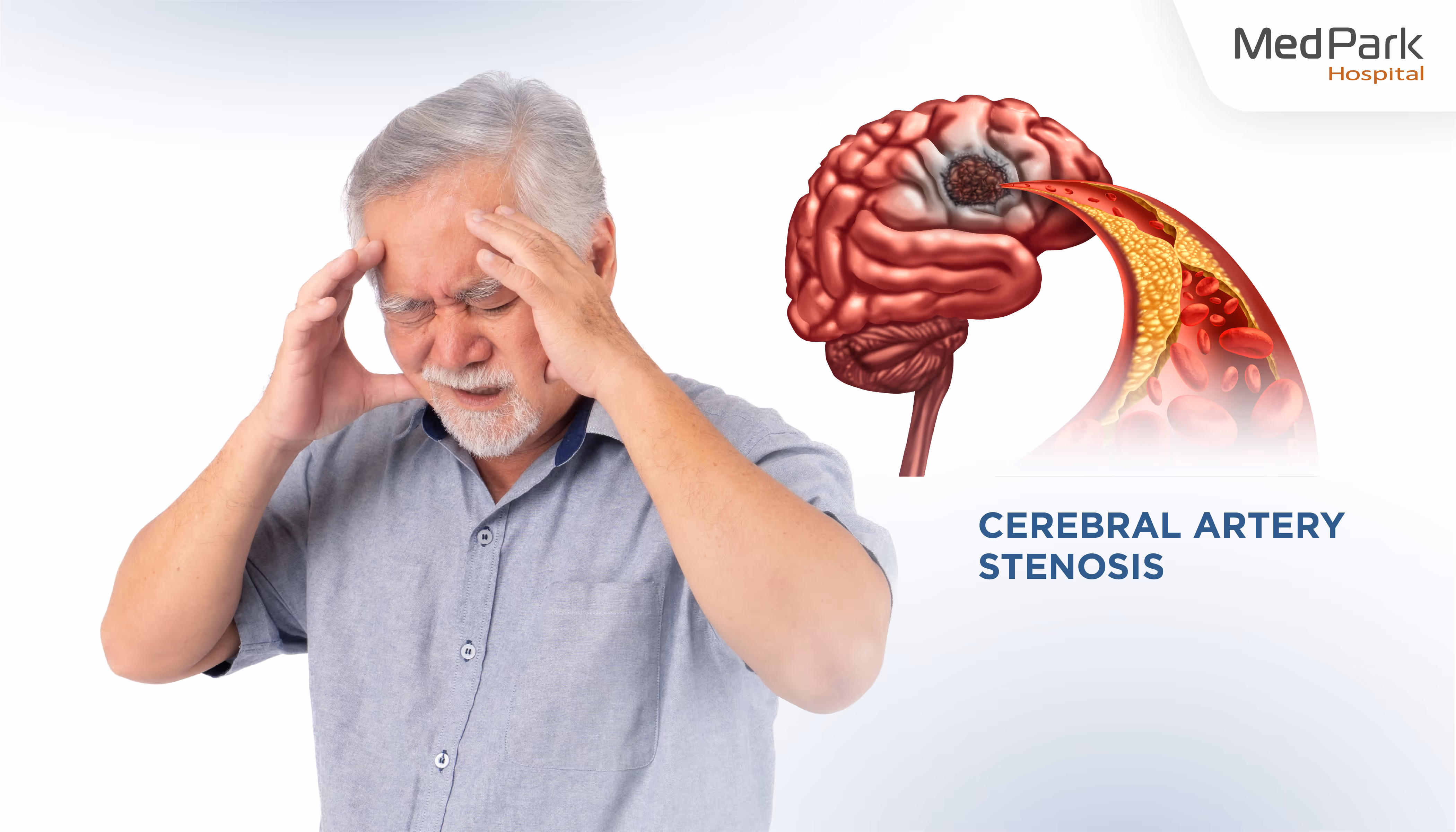
Cerebral angiogram is a medical procedure used to assess the risk of stroke by injecting contrast dye into a blood vessel and using X-ray imaging to detect cerebral artery stenosis, or an obstructed blood vessel in the brain that causes cerebral ischemia and paralysis. A cerebral angiogram assesses blood flow through the brain from the left and right carotid arteries, assisting in the detection of blood clots in the brain, brain bleeds, arteriovenous malformations, or aneurysms that cause severe headaches, limb weakness, Bell's palsy (one-sided facial drooping), unconsciousness, and even death. Cerebral angiogram lowers the risk of cerebral aneurysm, prevents disability, hemiplegia, and paralysis, and is a high-potential diagnostic test that yields accurate results and leads to timely treatment.
Why cerebral angiogram?
Typically, neurologists treat patients with intracranial stenosis caused by atherosclerosis or cerebral thrombosis who have had symptoms for less than 4.5 hours with thrombolytic or antiplatelet drugs to speed up the dissolution of the blood clot. However, in critical patients with large blood clots, those with contraindications to thrombolytic drugs, or those receiving the drug but whose blood vessels do not reopen, neurologists will render treatment by performing X-ray imaging guidance along with catheterization of the carotid artery to locate the blood clot in the brain and retrieve or aspirate it out to help restore the patency of cerebral blood vessels, allow blood to flow to the brain, and enable patients to overcome their life crisis.
What can be diagnosed with a cerebral angiogram?
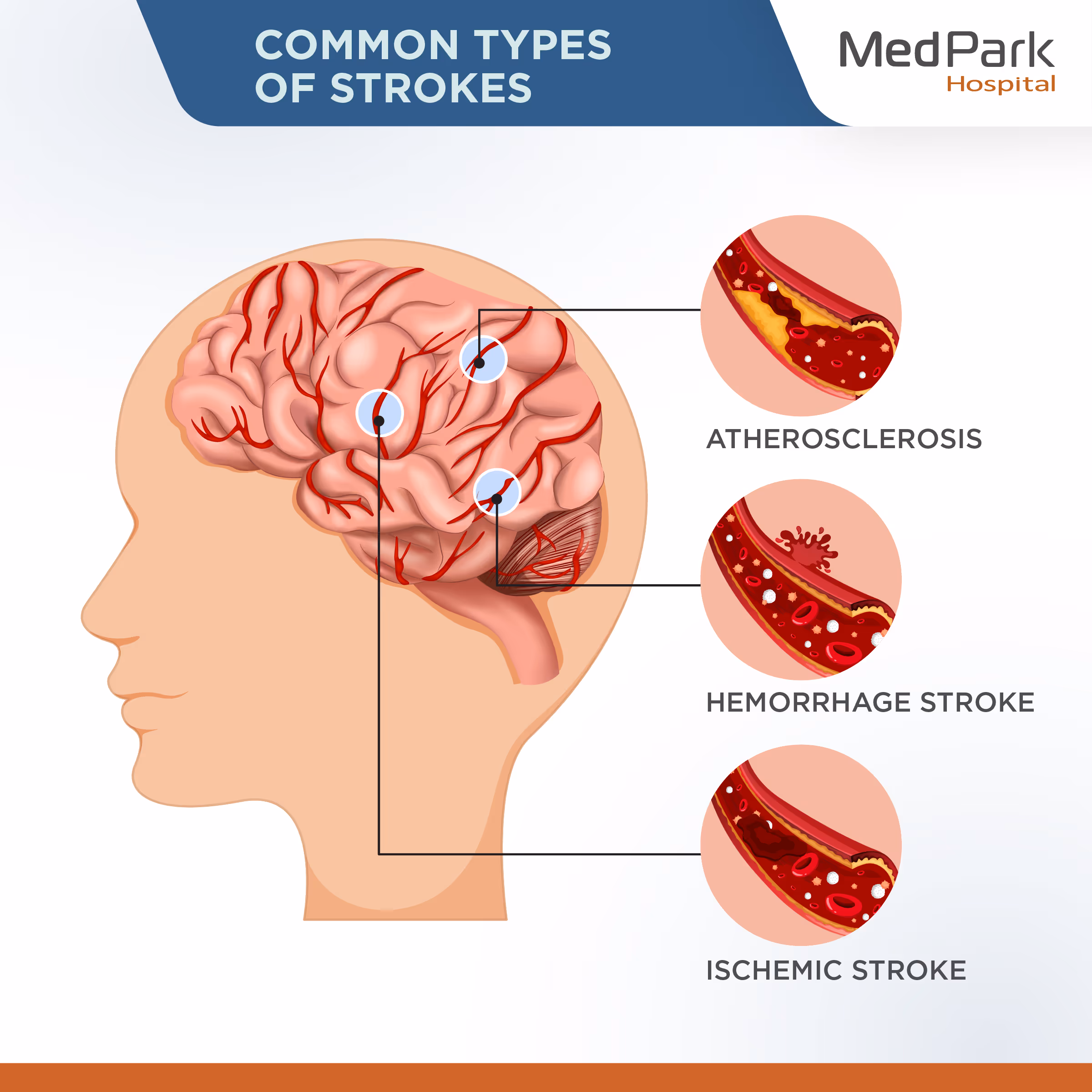
- Blood clot in the brain
- Intracranial stenosis (Cerebral artery stenosis)
- Ischemic stroke
- Intracerebral hemorrhage
- Hemorrhage stroke
- Brain aneurysm
- Atherosclerosis
- Vasculitis
- Vascular dissection
- Arteriovenous malformation (AVM)
- Dural arteriovenous fistula
What are the signs and symptoms of cerebrovascular disease?
- Acute severe headache, dizziness, sudden vertigo.
- Drooping mouth corners, drooping mouth, Bell's palsy.
- Slurred speech, difficulty speaking, stiff tongue, voice changes, communication problems
- Difficulty swallowing, choking, nausea, vomiting, and drowsiness.
- Sudden arm and leg weakness, acute numbness on one side of the face, hands, and feet.
- Sudden blurry vision, hazy vision, or double vision
- Loss of balance, foot drop, difficulty walking, or a staggering gait
- Peripheral cyanosis (blue hands and feet), body skin discoloration.
- Seizures, fainting, unconsciousness, and possibly death.
What is the cerebral angiogram procedure?
Cerebral angiograms at MedPark Hospital adhere to the gold standard for diagnosing and treating stroke and cerebrovascular disease, prioritizing the highest level of safety and the patient’s successful outcomes. Patients who undergo a mechanical thrombectomy for blood clot removal will require a one-night hospital stay for symptom monitoring before returning home if the postoperative conditions are satisfactory without any serious complications.
Pre-operative cerebral angiogram
- Patients with chronic diseases, personal medications, or a history of allergies to contrast dye must inform the neurologist in advance.
- Patients who take blood thinners such as aspirin, Plavix, or warfarin must inform the neurologist in advance.
- Before the examination, patients are subjected to X-rays, blood tests to assess kidney function and blood clotting, and electrocardiograms (for those over 35).
- For at least 6 hours before the examination, avoid eating, drinking, and smoking.
- The medical staff will inform the patients and their relatives about the examination procedures, including possible complications, and will have them sign a consent form before proceeding with the cerebral angiogram procedure.
Intraoperative cerebral angiogram
- The neurologist cleanses the catheter insertion site in the groin, wrist, or antecubital area before injecting local anesthesia to numb the area. The patient will feel slightly numb.
- For those without contraindications, the neurologist will insert a small catheter through the skin into the blood vessel and administer thrombolytic drugs intravenously (rt-PA) to quickly dissolve blood clots in the brain.
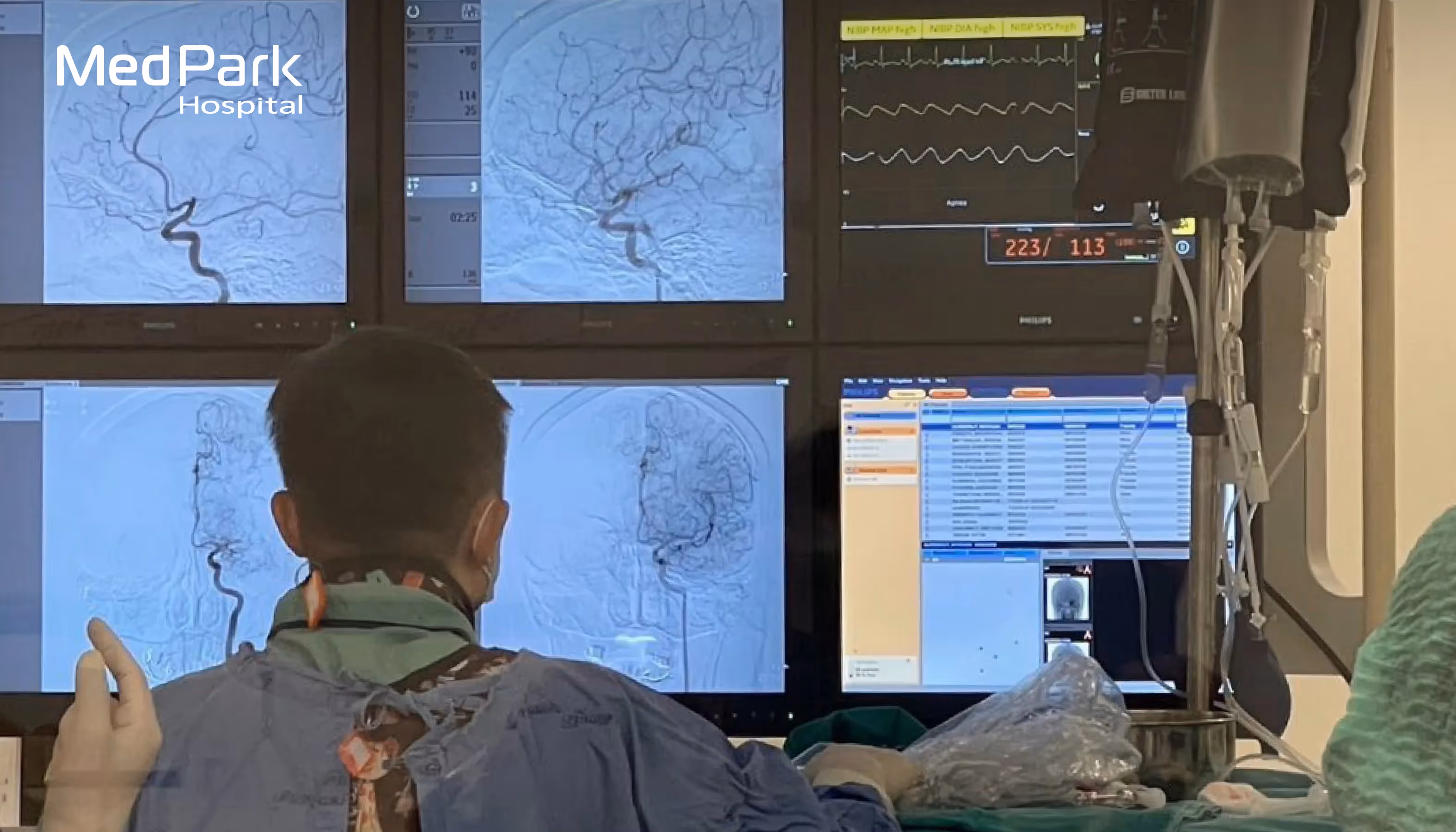
- The neurologist uses X-ray imaging guidance to thread a catheter along the artery, up to the carotid artery in the neck that connects to the cerebral vessels, to determine the location of the intracranial stenosis or occlusion.
- Then, the neurologist injects a contrast dye through the catheter, which displays the real-time X-ray image on an external screen, allowing the neurologist to clearly identify the location of the intracranial stenosis or occlusion, cerebral thrombosis, atherosclerosis, intracerebral hemorrhage, or other cerebrovascular abnormalities.
- If a blood clot is found, the neurologist will perform a mechanical thrombectomy by threading thrombectomy devices to retrieve or aspirate the blood clot from the brain, thereby opening the cerebral vessel and allowing blood to flow normally.
- When the examination and/or intervention is complete, the neurologist will withdraw the catheter from the artery and press on the puncture site for approximately 10-15 minutes to stop the bleeding. The patient will then move to the ward for further observation. Typically, a cerebral angiogram will take approximately 1-2 hours.
Post-operative cerebral angiogram
- Lie supine and avoid walking or bending the leg on the side where the procedure was performed for about 8 hours. If no abnormalities are found, the neurologist will allow the patient to return home.
- Take medication as prescribed by the neurologist to reduce pain and/or blood thinners to prevent blood clots.
- Drink 1-1.5 liters of clean water 2-3 days after the exam to help flush the contrast dye out of your system.
- If you experience numbness, coldness, pale skin, chest pain, palpitations, dizziness, fever, swelling, redness, bleeding at the puncture site, or are unable to lie flat, see a doctor right away.
What is the complication of a cerebral angiogram?
Common complications of a cerebral angiogram include bleeding, infection, blood vessel damage, and allergic reactions to the contrast dye. Severe complications from the cerebral angiogram that may occur include acute cerebral thrombosis, which may lead to paralysis. However, this serious complication occurred with a very low incidence rate of only 0.5%. Generally, a cerebral angiogram is considered a safe procedure, with a high success rate of up to 98.16% in assisting neurologists in identifying the location of intracranial stenosis or occlusion, which enables timely and effective intervention.

What are the benefits of a cerebral angiogram?
- Stroke screening: Stroke is the second leading cause of death worldwide. A stroke screening detects abnormalities in the brain's vessels, preventing brain aneurysms, intracranial stenosis, ruptures, or occlusions, which cause the brain to lose blood supply, brain cell death, and eventually death.
- Stroke risk assessment: It is estimated that one in every four people aged 25 and up has a stroke. A stroke screening assesses the risk of a stroke caused by a variety of risk factors in daily life, including high blood pressure, obesity, diabetes, smoking, and alcohol consumption.
- Non-surgical procedure: Cerebral angiogram uses X-ray imaging of the neck carotid arteries with an injection of a contrast dye into the cerebral vessels. The procedure is performed as an outpatient examination and does not require surgery or anesthesia. If the neurologist finds no abnormalities, the patient can return home the same day the examination is finished.
- Clear visualization: Cerebral angiogram is a diagnostic procedure that uses high-performance X-ray imaging and contrast material injected into the cerebral vessels. This enables the neurologist to see in more detail cerebral artery stenosis, rupture, blockage, aneurysm, brain hemorrhage, and other abnormalities than a CT scan or MRI.
- Combined diagnosis and treatment: During a cerebral angiogram, if a neurologist discovers cerebral artery stenosis or blockage, they will immediately retrieve or aspirate the blood clot from the brain. This makes a cerebral angiogram serve as both a diagnostic and therapeutic intervention at the same time. The procedure aids in the elimination of blood clots in the brain before symptoms become permanent, thereby lowering the mortality rate and the incidence of paralysis.

Cerebral Angiogram, MedPark Hospital
Neurology Clinic, MedPark Hospital, Bangkok, Thailand, is led by a team of neurologists and neurosurgeons specialized in the nervous system and brain disorders with extensive experience both domestically and internationally, ready to diagnose and treat common and complex neurological and brain disorders. including stroke/cerebrovascular disease, ischemic stroke, or acute severe headaches, using state-of-the-art technology and medical equipment with a gold standard treatment procedure to ensure accurate, rapid, and precise diagnosis, leading to timely and effective intervention; collaborate with multidisciplinary teams of neurology and brain clinical staff to provide 24-hour holistic care, ensuring the best handling of critical minutes where every minute is vital, allowing patients to recover quickly, free of complications, with a chance of cure, and enabling patients to resume their normal lives.