





Recognizing the Silent Danger of Retinal Disease, A Hidden Risk That Can Lead to Permanent Vision Loss
Each eye has only one retina, and once it is severely damaged, it cannot be repaired or replaced. As Thailand transitions into an aging society, combined with modern lifestyle factors such as poor diet and lack of exercise, and underlying conditions like diabetes, the incidence of retinal diseases is rising significantly.

In this Special Scoop, let us take a closer look at this silent retinal threat with Dr. Chanusnun Narongchai, an ophthalmologist specializing in retinal diseases at MedPark Hospital, who shares effective preventive measures and advanced, technology-driven treatments to restore sight and sustainable quality of life.
Retinal Diseases: If Left Untreated, Can Lead to Permanent Vision Loss
Many people often neglect eye health screenings because they do not experience any symptoms or they assume their vision issues are merely myopia or presbyopia. However, Dr. Chanusnun offered an insightful perspective on patients who come for consultations, categorizing them into two major groups:
- Asymptomatic patients often include adults over 40 who prioritize routine eye screenings, and a growing number of individuals in their 30s seeking preventive eye checkups.
- Symptomatic patients: This group spans a wide age range, from young adults in their 20s to older adults in their 70s and 80s. They often seek medical attention for symptoms such as blurred vision, floaters, or flashes of light. Being attentive to these symptoms is important, as many retinal conditions, if left untreated, can lead to permanent vision loss.
“The retina functions much like the extension of the brain and nervous system. It receives light and converts it into neural signals, allowing us to see images. If damage occurs, with prolonged cellular damage, recovery becomes increasingly difficult and complex,” said Dr. Chanusnun.
Critical Warning Signs: Visual Symptoms That Require Urgent Eye Care
To help you better assess your risk, the doctor highlights three common visual symptoms that may signal a serious underlying eye condition:
- Sudden vision loss: This may be caused by retinal detachment or an acute blockage of a major retinal artery. The condition is similar to an ischemic stroke, in which nerve cells are deprived of blood supply and may die rapidly if not treated in time.
- Black spots in the visual field, floating cobwebs, or seeing flashing lights in the eyes: These symptoms are common and are often caused by age-related vitreous degeneration. Such changes can lead to retinal thinning, holes, or tears. If left untreated, fluid can seep through these openings and eventually cause retinal detachment.
- Blurred central vision, faded colors, or distorted images: These symptoms are typically due to abnormalities in the macula. They typically develop gradually and may be associated with conditions such as diabetic macular edema, epiretinal membrane, or macular hemorrhage.
Three Common Retinal Diseases: A Serious Threat to Working-Age Adults and the Elderly
With modern lifestyles, retinal diseases are no longer limited to seniors. These conditions can generally be categorized into three main groups:
1. Age-Related Macular Degeneration (AMD)
This condition is closely linked to the natural aging process and is of two main types:
- Dry AMD: The most common form, characterized by slow progression. Vision gradually declines over time, allowing patients time to adapt and seek supportive care.
- Wet AMD: Less common but significantly more severe. It involves the growth of abnormal blood vessels beneath the retina. These fragile vessels can leak fluid or bleed, leading to rapid vision loss.
Many people believe that taking eye supplements can help slow age-related degeneration. However, Dr. Chanusnun provided important evidence-based medical insights on this topic.
“AREDS2-based supplements containing vitamin C, vitamin E, zinc, copper, lutein, and zeaxanthin are recommended only for certain patient groups and at specific stages of the disease, as determined by a doctor. It is important to consult an ophthalmologist before deciding to take them, as over-the-counter vitamins currently lack strong clinical evidence to support their efficacy in preventing this condition in healthy individuals. The most effective ways to reduce risk include avoiding smoking and keeping underlying conditions, such as diabetes, hypertension, and high cholesterol, well-controlled.”
2. Central Serous Chorioretinopathy (CSR): A Common Condition in Working Adults
Central serous chorioretinopathy (CSR) is a condition in which fluid builds up under the macula due to stress. It is increasingly common among office workers.
“We are seeing a steady rise in the number of working-age patients with CSR. The main risk factors are closely linked to lifestyle, particularly chronic work-related stress, sleep deprivation, inadequate rest, smoking, and the use of steroid medications. Patients typically present with blurred central vision, often describing a dark shadow in their field of vision, along with changes in color perception or visual distortion. Symptoms usually begin in one eye.” Dr. Chanusnun shared a case.
The condition is not severe enough to cause total blindness. In most cases, the fluid accumulation resolves on its own within 1–6 months, especially with strict lifestyle adjustments such as reducing stress and getting adequate rest. If it occurs only once and resolves quickly, it usually does not have a long-term impact on vision. However, if fluid persists for an extended period or recurs frequently, it may cause permanent damage to retinal cells, resulting in lasting visual impairment.
3. Diabetic Retinopathy
Diabetic retinopathy is a serious complication of diabetes, caused by persistently high blood sugar levels that damage the blood vessels in the eye. Over time, these vessels become fragile and leaky, leading to retinal swelling (macular edema) and the growth of abnormal new blood vessels. These fragile vessels can easily rupture, causing bleeding inside the eye and potentially significant vision loss.
“The most concerning aspect of diabetic retinopathy is that, in its early stages, patients often have no warning signs. Vision may seem normal until the disease has progressed to an advanced stage, before blurred vision begins to be noted,” Dr. Chanusnun added.

Retinal Diagnostic Innovations at MedPark Hospital
Accurate treatment begins with comprehensive screening. At MedPark Hospital, advanced diagnostic technologies enable clinicians to detect retinal abnormalities down to the cellular level with exceptional clarity.
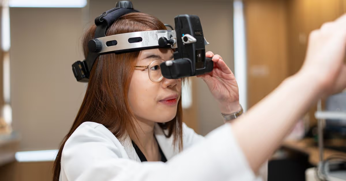
- Comprehensive Eye Exam: An eye examination including pupil dilation to assess the whole eye, from the anterior chamber and lens to intraocular pressure, optic nerve, and retinal vasculature in the posterior segment.
- Amsler Grid Test: A grid-patterned chart. Patients cover one eye at a time to test the center point. If the lines appear distorted or wavy, seek immediate medical attention.
- OCT (Optical Coherence Tomography): A high-resolution, light-based retinal scanning technology that provides detailed cross-sectional images of the retina, helping detect fibrosis, hemorrhage, or edema with high accuracy.
- Wide-field Fundus Photography: High-resolution, wide-field retinal imaging that enables rapid detection of retinal tears and other subtle peripheral lesions.

Restore Vision with 3 Modern Approaches to Treating Retinal Diseases
Ophthalmic technology has advanced significantly, reducing patient anxiety and enhancing the chances of restoring vision. Key treatment approaches include:
1. Intravitreal Injection
Intravitreal Injection is a standard treatment for retinal diseases such as wet AMD, diabetic macular edema, and retinal vein occlusion. Anti-VEGF agents are injected into the vitreous cavity to inhibit abnormal neovascularization and reduce retinal fluid and hemorrhage.
“Intravitreal injections are actually very straightforward. The procedure is quick, safe, and far less intimidating than many patients anticipate. Before the injection, the eye is carefully sterilized with an antiseptic solution, which may cause a mild stinging or tight sensation. A local anesthetic is then applied to ensure comfort. The injection itself takes only 1–2 seconds, and the needle is extremely fine. Many patients report little to no discomfort—at most, a brief sensation of pressure.”
“Intravitreal therapies have advanced significantly. Common medications include Avastin (bevacizumab), Lucentis (ranibizumab), Eylea (aflibercept), and Vabysmo (faricimab). This diverse range of highly effective options allows ophthalmologists to customize treatments with greater flexibility. If one medication becomes less effective, it can be adjusted or switched accordingly. Importantly, the newer generation of monoclonal antibodies has a longer duration of action, reducing the injection frequency for patients while achieving faster and more effective reduction of retinal fluid and hemorrhage,” Dr. Chanusnun clarified to ease patients’ concerns.
Typically, the initial phase requires monthly intravitreal injections for 3–6 months, depending on the disease and agent used. Afterward, dosing intervals are lengthened based on clinical response, with maintenance phases potentially extending up to every 3–6 months. The therapeutic effect generally manifests within 7 days after injection. Transient dark floaters may occur on the first day as the medication dissolves in the vitreous, which is entirely normal.
Laser therapy is a precisely targeted treatment modality, classified into panretinal photocoagulation (PRP) and focal laser (focal photocoagulation or retinopexy).
PRP is primarily indicated for advanced diabetic retinopathy with neovascularization. Its purpose is to suppress abnormal new vessel growth, reduce disease severity, and lower the risk of vessel rupture and subsequent intraocular hemorrhage. In addition to diabetic retinopathy, PRP may also be used in select cases of retinal vein occlusion or vasculitis, although these indications are less common.”
“Focal laser is a targeted treatment for retinal thinning, holes, or tears. The laser is applied around the affected area to prevent vitreous fluid from seeping beneath the retina and worsening retinal detachment. It functions as a protective dam against flooding,” Dr. Chanusnun explained.
The advanced multi-wavelength laser technology (red, yellow, and green) available at MedPark Hospital enables ophthalmologists to select the optimal wavelength based on specific pathology and lesion depth of a particular patient.
For advanced cases in which the disease has progressed to a severe stage, such as retinal detachment, fibrovascular traction, or non-clearing vitreous hemorrhage, vitrectomy is the definitive intervention. Recent advancements in surgical instrumentation have significantly enhanced procedural efficiency and speed, rendering modern vitreoretinal surgery much shorter and less daunting for patients.
“Modern surgical platforms use ultra–micro-gauge instruments, enabling sutureless procedures in many cases, including combined vitrectomy and cataract extraction. This minimally invasive approach shortens operative time, speeds recovery, reduces discomfort and foreign body sensation, and lowers the risk of suture-related inflammation,” added Dr. Chanasnan.
Beyond these advancements, gene therapy is an emerging field under clinical investigation for hereditary retinal dystrophies. It aims to slow the degeneration of photoreceptor cells at the DNA level, representing a new hope in ophthalmology worldwide.

Postoperative Care and Essential Guidelines for Visual Recovery After Retinal Surgery
Successful outcomes depend not only on the doctor’s expertise and advanced medical technology but also on the patient’s adherence to postoperative care instructions. Therefore, particular emphasis is placed on proper wound care and the correct use of prescribed medications.
“Avoid getting water in the eye during the initial recovery period to reduce the risk of infection. Use all prescribed eye drops as directed, including antibiotics, anti-inflammatory medications, and artificial tears. Wear sunglasses or protective eyewear to shield the eye from dust and other foreign particles that may cause irritation or inflammation. Some patients may need to avoid strenuous activities, such as heavy lifting or high-impact exercise, for approximately 2 weeks to 1 month to allow the eye to heal fully.”
For patients with underlying conditions, strict control of blood sugar and blood pressure is recommended both perioperatively and postoperatively to minimize the risk of disease progression (e.g., diabetic retinopathy) and prevent further retinal damage.
“I highly recommend that everyone aged 40 and older undergo routine annual comprehensive eye examinations, including retinal screenings. This age marks the onset of age-related ocular degeneration, which can manifest as epiretinal membranes, retinal thinning, or panretinal complications like diabetic retinopathy. The earlier these conditions are detected, the sooner we can initiate intervention, ultimately preventing and reducing the risk of irreversible vision loss,” Dr. Chanasnan concluded.
Because your delicate eyes deserve preventive care, begin your journey to better vision today at the Eye Care Center, MedPark Hospital.