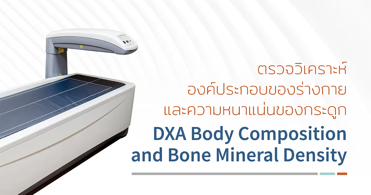
DXA คือ เครื่องตรวจวิเคราะห์ความหนาแน่นของกระดูก (Bone Mineral Density, BMD) รวมถึงโครงสร้างหรือคุณภาพภายในกระดูก (Trabecular Bone Score, TBS) เพื่อประเมินความเสี่ยงของการเกิดโรคกระดูกพรุน (Osteoporosis) และความเสี่ยงที่จะเกิดภาวะกระดูกหักอันเนื่องมาจากภาวะกระดูกพรุนในระยะเวลา 10 ปี นอกจากนี้เครื่อง DXA ยังสามารถตรวจวิเคราะห์องค์ประกอบต่าง ๆ ภายในร่างกาย ซึ่งได้แก่ มวลไขมัน มวลกล้ามเนื้อ และค่ามวลกระดูกเฉลี่ยทั้งร่างกาย ซึ่งถือเป็นการตรวจที่ได้รับการยอมรับจากทั่วโลก ว่ามีความถูกต้องและแม่นยำสูง ตรวจได้ง่าย รวดเร็ว โดยใช้ปริมาณรังสีน้อยมาก
องค์ประกอบที่เครื่อง DXA สามารถตรวจได้ มีดังนี้
- Lumbar spine การตรวจค่าความหนาแน่นของมวลกระดูกและความคุณภาพภายในกระดูกสันหลังส่วนเอว
- Femur การตรวจค่าความหนาแน่นของมวลกระดูกต้นขา
- VFA (vertebral fracture assessment) ตรวจดูแนวกระดูกหลังว่ามีการหัก การยุบตัวหรือไม่
- Body composition ตรวจวัดค่าไขมัน มวลกล้ามเนื้อ และค่ามวลกระดูกเฉลี่ยทั้งร่างกาย
การทำงานหลักๆ ของเครื่อง DXA SCAN
- ตรวจวิเคราะห์ความหนาแน่นของมวลกระดูก (BMD) ซึ่งโดยทั่วไปจะตรวจที่กระดูกสันหลังส่วนเอว (Lumbar spine) และกระดูกต้นขา (Femur) และอาจตรวจที่กระดูกบริเวณข้อมือ (Forearm) ในผู้ป่วยบางราย เช่น ผู้ป่วยที่มีระดับฮอร์โมนพาราไทรอยด์สูงมากกว่าปกติ (Hyperparathyroidism) หรือในคนไข้ที่ไม่สามารถตรวจที่บริเวณกระดูกสันหลังส่วนเอวหรือต้นขาได้ หรือในผู้ป่วยที่มีน้ำหนักมากกว่าปกติ จนเตียงไม่สามารถรับน้ำหนักได้ รวมถึงการตรวจโครงสร้างของกระดูกสันหลังส่วนเอว (TBS) เพื่อประเมินความเสี่ยงของการเกิดโรคกระดูกพรุน (osteoporosis) และความเสี่ยงในการเกิดภาวะกระดูกหักอันเนื่องมาจากภาวะกระดูกพรุนในระยะเวลา 10 ปี
- ตรวจวิเคราะห์องค์ประกอบต่าง ๆ ภายในร่างกาย ซึ่งได้แก่ มวลไขมัน มวลกล้ามเนื้อ และค่ามวลกระดูกเฉลี่ยทั้งตัว โดยเครื่องจะสามารถตรวจวิเคราะห์
- A. ปริมาณไขมันที่สะสมในร่างกาย โดยสามารถบอกทั้งปริมาณและสัดส่วน (percentage) เมื่อเทียบกับมวลกล้ามเนื้อเและกระดูก รวมถึงสามารถวิเคราะห์การกระจายตัวของไขมันว่าอยู่ที่ส่วนใด เช่น ไขมันสะสมบริเวณแขน ขา ลำตัว หรือสะโพก ซึ่งในปัจจุบันพบว่าข้อมูลการกระจายตัวของไขมันนั้น อาจมีความสำคัญมากกว่าปริมาณไขมันในร่างกาย โดยพบว่าการที่มีไขมันที่สะสมในช่องท้อง (Visceral fat) มากจะมีความเสี่ยงในการเกิดโรคระบบหัวใจและหลอดเลือด ความดันโลหิตสูง โรคเบาหวานชนิดดื้อต่ออินซูลิน ระดับไขมันในเลือดสูง ไขมันสะสมในตับ เป็นต้น
- B. มวลกล้ามเนื้อในร่างกาย โดยสามารถวิเคราะห์มวลกล้ามเนื้อรวม และแยกแยะมวลกล้ามเนื้อในส่วนต่างๆ ของร่างกาย เช่น มวลกล้ามเนื้อตามบริเวณแขน ขา ลำตัว ทั้งนี้เนื่องจากในคนสูงอายุ อาจมีมวลกล้ามเนื้อที่ลดลง ซึ่งอาจเป็นเหตุทำให้สุขภาพร่างกายโดยรวมไม่แข็งแรง หกล้มได้ง่าย หรือหากมีอาการเจ็บป่วย จะมีผลทำให้มีความเสี่ยงในการเกิดภาวะแทรกซ้อนสูงขึ้น หรือมีผลข้างเคียงจากยาสูงกว่าคนที่มีมวลกล้ามเนื้อปกติ นอกจากนี้จะมีประโยชน์ในคนที่ต้องการลดน้ำหนัก หากลดน้ำหนักผิดวิธี อาจมีผลให้มวลกล้ามเนื้อลดลง ซึ่งเป็นผลเสียมากกว่าผลดี หรือในนักกีฬาที่สร้างกล้ามเนื้อ (Body Builder) ข้อมูลกล้ามเนื้อในส่วนต่างๆ จะช่วยในการวางแผนการออกกำลังกายได้
นอกจากนี้ข้อมูลองค์ประกอบต่าง ๆ ภายในร่างกาย ทั้งมวลไขมันและกล้ามเนื้อ จะช่วยในการวางแผนทางโภชนาการ ทั้งในผู้ป่วยที่ต้องการลดน้ำหนัก ผู้ป่วยที่มีปัญหาเรื่องการทำงานของลำไส้ (Malabsorption) เป็นข้อมูลที่ช่วยในการปรับขนาดยาได้อย่างเหมาะสมในผู้ป่วยมะเร็ง การตรวจภาวะ HIV-associated lipodystrophy และในผู้ป่วยที่ต้องทำกายภาพบำบัด ก็จะช่วยในการปรับแผนการการรักษาได้เหมาะสมยิ่งขึ้น
การเตรียมตัวก่อนตรวจ
- สำหรับการตรวจ Bone Mineral Density (BMD)
- ไม่จำเป็นต้องงดน้ำและอาหาร
- การตรวจจะใช้เวลาประมาณ 15-20 นาที
- สำหรับการตรวจ Body Composition
- งดอาหารอย่างน้อย 4 ชั่วโมง แต่สามารถดื่มน้ำเปล่าได้
-
- งดเครื่องดื่มแอลกอฮอล์ อย่างน้อย 48 ชั่วโมง
- งดการออกกำลังกาย 12 ชั่วโมงก่อนการตรวจ
- การตรวจจะใช้เวลาประมาณ 20-30 นาที
- ควรเลื่อนการตรวจออกไป ในกรณีที่เคยได้รับการตรวจด้วยแบเรียม (Barium study) ในระยะเวลา 1 สัปดาห์ หรือได้รับการตรวจที่ใช้สารทึบรังสี หรือการตรวจทางเวชศาสตร์นิวเคลียร์ ในระยะเวลา 1-2 วัน
- ไม่แนะนำให้ทำในหญิงตั้งครรภ์ แม้ว่าการตรวจนี้มีปริมาณรังสีเพียงเล็กน้อย ดังนั้นหากสงสัยว่าตั้งครรภ์ หรือสงสัยว่าจะตั้งครรภ์ ควรเลื่อนการตรวจออกไปก่อน
ขั้นตอนการตรวจ
เปลี่ยนเสื้อผ้าเป็นชุดที่โรงพยาบาลเตรียมให้ ไม่ควรสวมชุดชั้นใน และเข้าห้องน้ำปัสสาวะให้เรียบร้อยก่อนรับการตรวจ หลังจากนั้นเจ้าหน้าที่จะให้ผู้ป่วยนอนบนเตียงและจัดท่าอย่างเหมาะสม ขณะตรวจให้อยู่นิ่ง ๆ ไม่ขยับ และสามารถหายใจได้ตามปกติ


