เลือกหัวข้อที่อ่าน
- ชนิดของโรคโพรงกระดูกสันหลังตีบแคบ
- อาการของโรคโพรงกระดูกสันหลังตีบแคบ
- สาเหตุของโรคโพรงกระดูกสันหลังตีบแคบ
- ความเสี่ยงในการเกิดโรคโพรงกระดูกสันหลังตีบแคบ
- แพทย์วินิจฉัยโรคโพรงกระดูกสันหลังตีบแคบอย่างไร?
- แพทย์รักษาโรคโพรงกระดูกสันหลังตีบแคบอย่างไร?
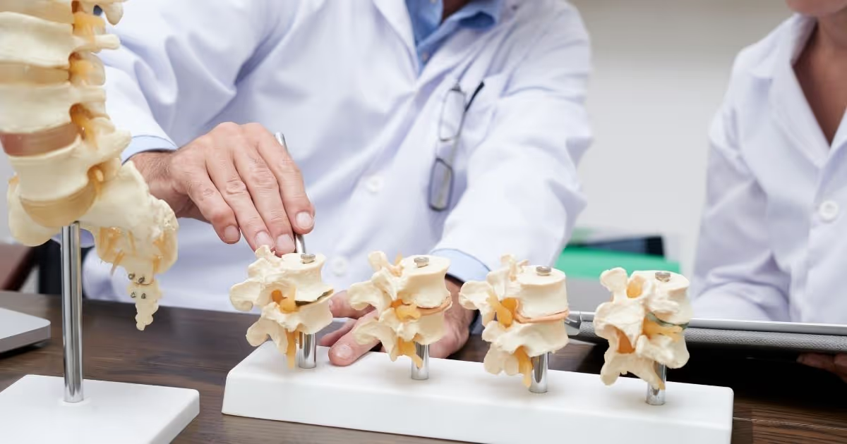
โรคโพรงกระดูกสันหลังตีบแคบ
โรคโพรงกระดูกสันหลังตีบแคบเป็นโรคที่เกิดจากความผิดปกติของโพรงกระดูกสันหลังที่แคบผิดปกติซึ่งส่งผลให้เส้นประสาทไขสันหลังถูกกดทับ โรคโพรงกระดูกสันหลังตีบแคบเกิดขึ้นบ่อยที่สุดในหลังส่วนล่าง ผู้ป่วยที่เป็นโรคโพรงกระดูกสันหลังตีบแคบอาจไม่แสดงอาการใด ๆ ในขณะที่ ผู้ป่วยบางรายอาจมีอาการปวด รู้สึกเสียวปลาบ มีอาการชาและกล้ามเนื้ออ่อนแรง และเมื่อเวลาผ่านไปนาน ๆ อาการอาจแย่ลงเรื่อย ๆ
โรคโพรงกระดูกสันหลังตีบแคบมักมีสาเหตุมาจากการเสื่อมสภาพของกระดูกสันหลังที่เกี่ยวเนื่องกับโรคข้อเข่าเสื่อมนกรณีที่อาการของโรคโพรงกระดูกสันหลังตีบแคบร้ายแรง แพทย์อาจแนะนําให้เข้ารับการผ่าตัดเพื่อเพิ่มพื้นที่ให้เส้นประสาทไขสันหลังหรือเส้นประสาท
ชนิดของโรคโพรงกระดูกสันหลังตีบแคบ
แพทย์จะแบ่งชนิดของโรคโพรงกระดูกสันหลังตีบแคบตามตำแหน่งที่เกิดโรคบนแนวกระดูกสันหลัง ทั้งนี้ ผู้ป่วยบางรายอาจมีโรคโพรงกระดูกสันหลังตีบแคบหลายชนิดด้วยกัน โรคโพรงกระดูกสันหลังตีบแคบมีด้วยกัน 2 ชนิดหลักได้แก่:
- กระดูกต้นคอตีบ (Cervical Stenosi)ซึ่งจะเกิดขึ้นในกระดูกสันหลังบริเวณคอ
- โพรงประสาทส่วนเอวตีบแคบ (Lumbar stenosis) ซึ่งจะเกิดขึ้นในกระดูกสันหลังบริเวณหลังส่วนล่าง ซึ่งโรคโพรงกระดูกสันหลังตีบแคบชนิดนี้พบมากที่สุดในกลุ่มโรคโพรงกระดูกสันหลังตีบแคบด้วยกัน
อาการของโรคโพรงกระดูกสันหลังตีบแคบ
จากหลายกรณีพบว่า โรคโพรงกระดูกสันหลังตีบแคบมักพบได้จากการตรวจด้วยเครื่องสร้างภาพด้วยสนามแม่เหล็กไฟฟ้าหรือการตรวจด้วยเครื่องเอกซเรย์คอมพิวเตอร์ ทั้งนี้ ผู้ป่วยหลายรายอาจไม่แสดงอาการแม้จะมีโรคโพรงกระดูกสันหลังตีบแคบ โดยโรคมักจะเริ่มค่อย ๆ เกิดและแย่ลงไปเรื่อย อาการของโรคโพรงกระดูกสันหลังตีบแคบอาจแตกต่างกันไปในผู้ป่วยแต่ละราย โดยขึ้นอยู่กับตําแหน่งของโรคและเส้นประสาทที่ได้รับผลกระทบ
เมื่อเกิดขึ้นกับกระดูกสันหลังส่วนคอ:
- จะเกิดอาการชา หรือรู้สึกเสียวปลาบตามมือ แขน เท้า หรือขา
- รู้สึกมือ แขน เท้าและขาอ่อนแรง
- มีปัญหาเกี่ยวกับการเดินและการทรงตัว
- มีอาการปวดคอ
- ในกรณีที่รุนแรง ผู้ป่วยอาจรู้สึกถึงความผิดปกติในลำไส้และกระเพาะปัสสาวะ
เมื่อเกิดขึ้นกับกระดูกสันหลังส่วนเอว:
- รู้สึกชาหรือเสียวปลาบในเท้าทั้งสองข้างหรือข้างใดข้างหนึ่ง
- เท้าหรือขารู้สึกอ่อนแรง
- มีอาการปวดหรือตะคริวในขาข้างใดข้างหนึ่งหรือทั้งสองข้าง จะเกิดขึ้นเมื่อต้องยืนหรือเดินนาน ๆ โดยจะหายได้เองเมื่อโน้มตัวไปข้างหน้าหรือนั่งลง
- มีอาการปวดหลัง
- เดินได้แค่ระยะใกล้ เช่น 50-150 เมตร แล้วต้องนั่งพักซักครู่ ก่อนจะเริ่มเดินได้อีกครั้ง
สาเหตุของโรคโพรงกระดูกสันหลังตีบแคบ
โดยปกติแล้ว กระดูกสันหลังของมนุษย์จะทอดยาวจากคอลงไปยังหลังส่วนล่าง โดยกระดูกสันหลังจะมีช่องบรรจุไขสันหลังซึ่งช่วยปกป้องเส้นประสาทไขสันหลัง (เส้นประสาท) เอาไว้ ทั้งนี้ บางคนอาจเกิดมาพร้อมกับช่องบรรจุไขสันหลังขนาดเล็ก แต่โรคโพรงกระดูกสันหลังตีบแคบส่วนใหญ่เกิดขึ้นเมื่อโพรงกระดูกสันหลังแคบจนผิดปกติแล้ว
สาเหตุของโรคโพรงกระดูกสันหลังตีบแคบ ได้แก่:
- กระดูกโตผิดปกติความเสื่อมอันเกิดจากโรคข้อเข่าเสื่อมในกระดูกสันหลังอาจกระตุ้นให้เกิดภาวะกระดูกงอกในช่องบรรจุไขสันหลัง ซึ่งเป็นโรคที่มักเกิดในผู้ใหญ่ และเป็นเหตุให้เกิดกระดูกโตผิดปกติในกระดูกสันหลังได้อีกด้วย
- หมอนรองกระดูกเคลื่อนทับเส้นประสาท (Herniated Discs) เป็นภาวะที่หมอนรองกระดูกที่ทําหน้าที่กันกระแทกระหว่างกระดูกสันหลังมีแนวโน้มที่จะแห้งไปตามอายุ ทั้งนี้ เมื่อเกิดรอยแตกด้านนอกอาจช่วยให้เนื้อเยื่อด้านในของหมอนรองกระดูกโผล่ออกมาและกดทับเส้นประสาทไขสันหลังหรือเส้นประสาท
- เอ็นยึดกระดูกหนาผิดปกติเป็นภาวะที่เส้นเอ็นที่ยึดกระดูกสันหลังไว้ด้วยกันเกิดหนาตัวและเกิดเป็นพังผืดขึ้น ซึ่งเอ็นเหล่านี้จะยื่นเข้าไปในช่องบรรจุไขสันหลัง
- เนื้องอกในไขสันหลัง เกิดขึ้นภายในเยื่อหุ้มเส้นประสาทกระดูกสันหลัง หรือในพื้นที่ระหว่างไขสันหลังและกระดูกสันหลัง โดยเนื้องอกสามารถพบได้จากการตรวจวินิฉัยด้วยภาพถ่ายกระดูกสันหลัง พร้อมใช้วิธีตรวจด้วยเครื่องสร้างภาพด้วยสนามแม่เหล็กไฟฟ้า (MRI) และการแสดงภาพด้วยคลื่นแม่เหล็กและคลื่นเสียงความถี่สูง (CT) ร่วมด้วย
- การบาดเจ็บบริเวณกระดูกสันหลัง อุบัติเหตุทางรถยนต์ และการบาดเจ็บอื่น ๆ อาจทําให้กระดูกสันหลังเคลื่อนหรือหัก กระดูกเคลื่อนอันเกิดจากกระดูกสันหลังหักอาจก่อความเสียหายให้กับเนื้อเยื่อในช่องกระดูกสันหลัง อาการบวมของเนื้อเยื่อใกล้เคียงที่เกิดขึ้นทันทีหลังการผ่าตัดอาจกดทับเส้นประสาทหรือเส้นประสาทกระดูกสันหลัง
ความเสี่ยงในการเกิดโรคโพรงกระดูกสันหลังตีบแคบ
ผู้ป่วยที่เป็นโรคโพรงกระดูกสันหลังตีบแคบส่วนใหญ่มีอายุมากกว่า 50 ปีขึ้นไป แม้การเสื่อมสภาพตามวัยอาจทําให้เกิดโรคโพรงกระดูกสันหลังตีบแคบในผู้ที่มีอายุน้อย แต่ก็ยังมีสาเหตุอื่น ๆ ในการเกิดโรคโพรงกระดูกสันหลังตีบแคบด้วย โดยรวมถึง การบาดเจ็บ กระดูกสันหลังผิดรูปแต่กำเนิด เช่น กระดูกสันหลังคด และโรคทางพันธุกรรมที่มีผลต่อการพัฒนากระดูกและกล้ามเนื้อทั่วร่างกาย การตรวจกระดูกสันหลังด้วยการตรวจวินิฉัยด้วยภาพถ่ายสามารถระบุและแยกสาเหตุของโรคได้
ภาวะแทรกซ้อน
โรคโพรงกระดูกสันหลังตีบแคบน้อยชนิดเป็นชนิดรุนแรง และหากไม้ได้รับการรักษาอาจทำให้เกิดความรุนแรงมากขึ้นกว่าเดิมและไม่สามารถรักษาให้หายได้ โดยอาการทำให้เกิดอาการเหล่านี้หนักขึ้นหรือถาวร เช่น
- มีอาการชา
- กล้ามเนื้ออ่อนแรง
- มีปัญหาในการทรงตัว
- ควบคุมการขับถ่ายอุจจาระและปัสสาวะไม่ได้
- อัมพาต
แพทย์วินิจฉัยโรคโพรงกระดูกสันหลังตีบแคบอย่างไร
เพื่อวินิจฉัยโรคโพรงกระดูกสันหลังตีบแคบ แพทย์อาจสอบถามอาการผู้ป่วยก่อน โดยมีการซักประวัติคนไข้ และอาจตรวจร่างกายร่วมด้วย โดยแพทย์ใช้วิธีการตรวจวินิฉัยด้วยภาพถ่ายร่วมด้วยเพื่อช่วยระบุสาเหตุของอาการ
การตรวจวินิฉัยด้วยภาพถ่าย
การตรวจวินิฉัยด้วยภาพถ่ายที่แพทย์ใช้เพื่อการวินิจฉัย ได้แก่:
- การเอกซเรย์ (X-Ray)การเอกซเรย์ช่วยให้แพทย์เห็นการเปลี่ยนแปลงของกระดูก เช่น ภาวะกระดูกงอกที่อาจทำให้พื้นที่ในช่องบรรจุไขสันหลังแคบลง อย่างไรก็ตาม การทำการเอกซเรย์แต่ละครั้ง ผู้ป่วยจะได้รับรังสีเอ็กซ์ไปด้วย
- การตรวจด้วยเครื่องสร้างภาพด้วยสนามแม่เหล็กไฟฟ้า (MRI)เป็นการตรวจแบบใช้แม่เหล็กที่ทรงพลังและคลื่นวิทยุเพื่อสร้างภาพตัดขวางของกระดูกสันหลัง การตรวจนี้จะช่วยให้แพทย์ตรวจสอบความเสียหายของหมอนรองกระดูกและเอ็นยึดกระดูก และตรวจดูว่ามีเนื้องอกหรือไม่ ที่สําคัญที่สุด การตรวจนี้ช่วยให้แพทย์สามารถระบุตำแหน่งของเส้นประสาทในไขสันหลังที่ถูกกดทับได้
- การตรวจวินิจฉัยโรคด้วยเครื่องเอกซเรย์คอมพิวเตอร์หรือการฉีดสีเข้าไขสันหลังแล้วถ่ายภาพรังสี หากใช้การตรวจด้วยเครื่องสร้างภาพด้วยสนามแม่เหล็กไฟฟ้าไม่ได้ แพทย์อาจแนะนำวิธีการตรวจวินิจฉัยโรคด้วยเครื่องเอกซเรย์คอมพิวเตอร์ ซึ่งจะใช้การตรวจวินิฉัยด้วยภาพถ่ายร่วมด้วย โดยเป็นการถ่ายจากมุมต่างกันเพื่อสร้างภาพตัดขวางของร่างกายแบบละเอียด ทั้งนี้ ในการฉีดสีเข้าไขสันหลังแล้วถ่ายภาพรังสีนั้น แพทย์จะทำการตรวจวินิจฉัยโรคด้วยเครื่องเอกซเรย์คอมพิวเตอร์หลังจากการฉีดสีเข้าไขสันหลังแล้ว โดยสีที่ฉีดเข้าไปทำให้เห็นไขสันหลัง และเส้นประสาท และช่วยให้แพทย์สามารถระบุหมอนรองกระดูกเคลื่อนทับเส้นประสาท ภาวะกระดูกงอกและเนื้องอกได้ ถ้ามี
แพทย์รักษาโรคโพรงกระดูกสันหลังตีบแคบอย่างไร?
การผ่าตัดกระดูกสันหลัง
การรักษาโรคโพรงกระดูกสันหลังตีบแคบขึ้นอยู่กับตําแหน่งของโรคและความรุนแรงของอาการ
การใช้ยารักษา
แพทย์สั่งยาเหล่านี้เพื่อรักษาโรคโพรงกระดูกสันหลังตีบแคบ:
- ยาบรรเทาอาการปวดแพทย์อาจสั่งยาแก้ปวดให้ ได้แก่ ไอบูโพรเฟน นาพร็อกเซน และอะเซตามิโนเฟน ให้ผู้ป่วยเพื่อช่วยบรรเทาอาการปวดอันเกิดจากโรคโพรงกระดูกสันหลังตีบแคบชั่วคราว ทั้งนี้ ยาเหล่านี้ควรใช้ในระยะสั้นเท่านั้น เนื่องจากยังมีงานวิจัยเกี่ยวกับประโยชน์จากการใช้ยาเหล่านี้ในระยะยาวยังมีไม่เพียงพอ
- ยารักษาโรคซึมเศร้าแพทย์อาจสั่งยาต้านโรคซึมเศร้ากลุ่มไตรไซคลิก เช่น อะมิทริปไทลีน เพื่อช่วยบรรเทาอาการปวดเรื้อรังในเวลากลางคืน
- ยากันชักแพทย์อาจสั่งยากันชักบางชนิด เช่น กาบาเพนตินและพรีกาบาลิน สามารถใช้ลดอาการปวดที่เกิดจากเส้นประสาทถูกทำลาย
- สารโอปิออยด์ เป็นยาที่ประกอบด้วยสารเสพติดจากโคเคน เช่น ออกซิโคโดนและไฮโดรโคโดน เพื่อช่วยบรรเทาปวดชั่วคราว นอกจากนี้ แพทย์อาจนำเอาสารโอปิออยด์มาใช้ในการรักษาระยะยาวและภายใต้การดูแลของแพทย์อย่างใกล้ชิด ทั้งนี้ ยาเหล่านี้มีความเสี่ยงในการก่อผลข้างเคียงที่รุนแรง รวมทั้ง การติดยาหรือการขาดยาไม่ได้
กายภาพบําบัด
โดยปกติแล้ว ผู้ป่วยที่เป็นโรคโพรงกระดูกสันหลังตีบแคบจะขยับตัวหรือทำกิจกรรมเคลื่อนไหวน้อยลงเพื่อลดอาการปวด ซึ่งจะทำให้กล้ามเนื้ออ่อนแรงและเกิดอาการปวดมากขึ้น ทั้งนี้ นักกายภาพบําบัดสามารถช่วยฝึกให้ผู้ป่วยออกกำลังกายเพื่อ:
- สร้างความแข็งแรงและแข็งแกร่งให้ร่างกาย
- ช่วยรักษาความยืดหยุ่นและแข็งแรงของกระดูกสันหลัง
- ช่วยให้การทรวงตัวดีขึ้น
การฉีดสเตียรอยด์
รากประสาทอาจเกิดการระคายเคืองและบวมบริเวณที่ฉีดได้ การฉีดยาสเตียรอยด์ (ยาคอร์ติโคสเตียรอยด์) ในบริเวณที่เกิดโรคไม่ได้ช่วยรักษาโรคโพรงกระดูกสันหลังตีบโดยตรง แต่จะช่วยลดการอักเสบและบรรเทาอาการปวดเท่านั้น อย่างไรก็ตาม การฉีดสเตียรอยด์ไม่ได้เหมาะกับผู้ป่วยทุกราย และการฉีดเตียรอยด์ซ้ําอาจทำให้กระดูกในบริเวณใกล้เคียงและเนื้อเยื่อเกี่ยวพันอ่อนแอ ดังนั้น แพทย์อาจสั่งให้ใช้วิธีการฉีดสเตียรอยด์ได้ไม่กี่ครั้งต่อปีเท่านั้น
การผ่าตัดแก้ไขการกดทับเส้นประสาท
ด้วยวิธีนี้ แพทย์จะใช้เครื่องมือคล้ายเข็มเพื่อตัดเอาส่วนเส้นเอ็นหนาผิดปกติบริเวณด้านหลังของแนวกระดูกสันหลังออก เพื่อเพิ่มพื้นที่ช่องบรรจุไขสันหลังและลดอันตรายและบาดเจ็บที่เส้นประสาทอันเกิดจากการกดทับ ทั้งนี้ แพทย์จะใช้วิธีการผ่าตัดนี้เฉพาะกับผู้ป่วยที่เป็นโรคโพรงกระดูกสันหลังตีบแคบและเส้นเอ็นหนาผิดปกติเท่านั้น ทั้งนี้ การผ่าตัดนี้เรียกว่าการแก้ไขการกดทับที่กระดูกสันหลังส่วนเอวผ่านผิวหนังโดยใช้ภาพถ่ายทางรังสี (PILD) หรือ การผ่าตัดกระดูกสันหลังส่วนเอวแบบแผลเล็ก (MILD) แต่โดยส่วนใหญ่แพทย์มักใช้ การแก้ไขการกดทับที่กระดูกสันหลังส่วนเอวผ่านผิวหนังโดยใช้ภาพถ่ายทางรังสี เนื่องจากสามารถทำได้โดยไม่ต้องใช้ยาระงับความรู้สึกทั่วไป นอกจากนี้ แพทย์ยังเลือกใช้วิธีนี้กับผู้ป่วยบางรายที่อาจเกิดปัญหาสุขภาพจากการผ่าตัด
ศัลยกรรมผ่าตัด
แพทย์อาจใช้วิธีทำศัลยกรรมผ่าตัดหากการรักษาอื่น ๆ ไม่ได้ผลหรืออาการรุนแรง โดยเป้าหมายของการผ่าตัดคือการบรรเทาอาการกดทับในไขสันหลังหรือรากประสาทโดยการสร้างพื้นที่มากขึ้นภายในช่องบรรจุไขสันหลัง การผ่าตัดแก้ไขการกดทับเส้นประสาทเป็นวิธีที่นำมาใช้บ่อยเพื่อรักษาโรคโพรงกระดูกสันหลังตีบแคบ
การผ่าตัดเพื่อรักษาโรคโพรงกระดูกสันหลังตีบแคบ ได้แก่:
- การผ่าตัดกระดูกสันหลังแบบลามิเนกโตมีจะเป็นการผ่าตัดยกกระดูกลามินาออกจากกระดูกสันหลังที่กดทับ ทั้งนี้ การผ่าตัดกระดูกสันหลังแบบลามิเนกโตมี บางครั้งเรียกว่า การผ่าตัดแก้ไขการกดทับเส้นประสาท เพราะช่วยลดการกดทับของเส้นประสาทโดยการสร้างพื้นว่างรอบ ๆ ในบางกรณี แพทย์อาจใช้เครื่องมือช่วยยึดกระดูกสันหลังหรือที่เรียกว่า การเชื่อมข้อกระดูกสันหลังเพื่อรักษาความแข็งแรงของกระดูกสันหลัง
- การผ่าตัดเปิดบางส่วนของกระดูกลามินาออก (Laminotomy)ด้วยวิธีการนี้ แพทย์จะผ่าตัดเอาบางส่วนของกระดูกลามินาออกโดยเจาะรูให้ใหญ่พอที่จะลดแรงกดในจุดใดจุดหนึ่งลงได้
- การผ่าตัดขยายโพรงกระดูกสันหลัง (Laminoplasty) แพทย์จะใช้วิธีนี้ผ่าตัดกระดูกสันหลังบริเวณคอเท่านั้น เพื่อเปิดพื้นที่ภายในช่องบรรจุไขสันหลังโดยการสร้างฝาเล็ก ๆ บนกระดูกลามินา และแพทย์จะใช้โลหะยึดเชื่อมช่องว่างในกระดูกสันหลังที่เปิดออก
- การศัลยกรรมผ่าตัดผ่านกล้องด้วยวิธีการนี้ แพทย์จะผ่าตัดเอากระดูกหรือกระดูกลามินาออกเพื่อลดความเสียหายต่อเนื้อเยื่อดีที่อยู่ใกล้เคียง โดยจะทำให้ไม่จำเป็นต้องผ่าเชื่อมกระดูก
แม้การผ่าเชื่อมกระดูกเป็นวิธีที่ช่วยเสริมความแข็งแกร่งของกระดูกสันหลังและลดอาการปวด อย่างไรก็ตาม หากผู้ป่วยไม่ใช้วิธีผ่าเชื่อมกระดูกก็จะลดความเสี่ยงในการเกิดอาการอื่น ๆ เช่น อาการปวดหลังผ่าตัด และการอักเสบหลังผ่าตัด และโรคต่าง ๆ ที่อาจเกิดขึ้นกับพื้นที่ใกล้เคียงกระดูกสันหลัง นอกจากการลดความจําเป็นในการผ่าเชื่อมกระดูกแล้ว การศัลยกรรมผ่าตัดผ่านกล้องรวมถึงการผ่าตัดต่าง ๆ อาจทำให้ผู้ป่วยฟื้นตัวได้เร็วขึ้น
ทั้งนี้ การผ่าตัดเพื่อเพิ่มพื้นที่ในกระดูกสันหลังช่วยลดอาการของโรคโพรงกระดูกสันหลังตีบแคบ แต่ในบางกรณี ผู้ป่วยอาจไม่ได้มีอาการดีขึ้นหรือแย่ลงหลังการผ่าตัดแล้ว ทั้งนี้ หลังการผ่านตัด ผู้ป่วยอาจเสี่ยงได้รับผลกระทบอื่น ๆ ได้แก่ การติดเชื้อ การฉีกขาดในเยื่อหุ้มเซลล์ที่คลุมเส้นประสาทไขสันหลัง เกิดลิ่มเลือดในหลอดเลือดดําที่ขา และเกิดการเสื่อมของระบบประสาท